A look at the true nature of the zombie brain
Even if you've never seen a zombie movie or television show, you could identify an undead ghoul if you saw one. With their endless wandering, lumbering gait, insatiable hunger, antisocial behavior, and apparently memory-less existence, zombies are the walking nightmares of our deepest fears. What do these characteristic behaviors reveal about the inner workings of the zombie mind? Could we diagnose zombism as a neurological condition by studying their behavior? In Do Zombies Dream of Undead Sheep?, neuroscientists and zombie enthusiasts Timothy Verstynen and Bradley Voytek apply their neuro-know-how to dissect the puzzle of what has happened to the zombie brain to make the undead act differently than their human prey.
Combining tongue-in-cheek analysis with modern neuroscientific principles, Verstynen and Voytek show how zombism can be understood in terms of current knowledge regarding how the brain works. In each chapter, the authors draw on zombie popular culture and identify a characteristic zombie behavior that can be explained using neuroanatomy, neurophysiology, and brain-behavior relationships. Through this exploration they shed light on fundamental neuroscientific questions such as: How does the brain function during sleeping and waking? What neural systems control movement? What is the nature of sensory perception?
Walking an ingenious line between seriousness and satire, Do Zombies Dream of Undead Sheep? leverages the popularity of zombie culture in order to give readers a solid foundation in neuroscience.
eBook - ePub
Do Zombies Dream of Undead Sheep?
A Neuroscientific View of the Zombie Brain
- 272 pages
- English
- ePUB (mobile friendly)
- Available on iOS & Android
eBook - ePub
Do Zombies Dream of Undead Sheep?
A Neuroscientific View of the Zombie Brain
About this book
Trusted by 375,005 students
Access to over 1.5 million titles for a fair monthly price.
Study more efficiently using our study tools.
Information
Publisher
Princeton University PressYear
2014Print ISBN
9780691173153
9780691157283
eBook ISBN
9781400851928

GRAY’S (UNDEAD) ANATOMY
With savages, the weak in body or mind are soon eliminated; and those that survive commonly exhibit a vigorous state of health.
—Charles Darwin, The Descent of Man
You are about to read a book about the zombie brain. Just think about that for a minute. Let the thought really soak in. Reflect on the decisions you’ve made in your life that led you to this point.
Now let’s get a bit meta for a moment and think about all of that thinking and reflecting you just did. First, you read some words that we wrote via a semi-creative process. You understood those words and they changed your behavior. You reflected on your life by some internal memory recollection process. Perhaps you even thought about what decisions led us to the point of writing this book in the first place.
This amalgamation of thoughts, memories, and emotions that you just experienced, and will keep experiencing while reading this book, are all the result of a never-ending symphony of electrochemical processes in your brain. Each step of thinking that you just performed, from seeing the printed letters on the page to following the linguistic requests that we asked of you by pulling up the memories of the past, is performed by little networks of neurons distributed throughout that gray matter sandwiched inside your skull.
As neuroscientists, the fact that we can do all of that “thinking” is completely amazing. But what if you couldn’t do any of that? Or what if you could do some of those things, but could feel no emotions about them? Or what if you could feel emotion, but had no memory?
The study of neuroscience isn’t just about tissues and neurons and signals; it also has strong philosophical, computational, and psychological roots. It is a very difficult, sometimes wonderful, but often frustrating, problem.
Which is how we got to this point. As we said in the introduction, this is a book by a couple of scientists who also happen to be zombie movie nerds.
Our goal for this little thought experiment is to understand what has happened to the walking dead that has changed them from normal human beings to so-called “mindless walking corpses.”1 To do this we need to understand how the brain gives rise to behavior, in both humans and zombies. Which means we first have to understand exactly what the brain is.
But before we can get knee-deep in zombie gray matter, let us take a step back and look at the little three-pound piece of tissue sandwiched between your ears.
NEUROSCIENCE WITHOUT BRAIN SCANNERS
In this chapter and those that follow, we will attempt to link features of zombie behaviors to the various parts of the brain by adopting a classical forensic neurology approach.
What do we mean by this?
Classic neurology was the original scientific method for studying the brain before we had big machines to take pictures inside the living skull. Neurology is mainly focused on understanding why certain things go wrong in the brain to cause a patient’s symptoms, but along the way it has learned a lot about how the healthy brain works too. When neurology began in the mid-1800s, doctors had to deduce how the brain works by simply observing the behaviors of people and animals. This is a delicate art that involves making deductions about the brain by carefully detailing your subject’s behavior. But it didn’t just start with the advent of neurology in the nineteenth century. In fact, this form of investigation has been going on for centuries.
Indeed, while we tend to think of neuroscience (the empirical study of the healthy brain, as opposed to neurology, which is the medical branch dealing with brain disorders) as a “modern” scientific endeavor, some of the first experimental research linking the brain and nerves to behavior came from experiments and demonstrations by the Roman physician Claudius Galen, sometime between 150 and 190 CE.
Keep in mind that we’re talking about a time nearly 2000 years before brain imaging, well before Dr. House could just send his patients to an MRI to see how healthy their brains were. Back then, physicians and scientists had to do a lot with very little information. They had to get creative. This meant that they tried a lot of things; some worked and some didn’t. But sometimes they learned something new that would add just a bit more to what little was known about the brain.
For example, in a famous experiment on a living pig, Galen was trying to trace out the nerves involved in breath control when he accidentally cut the recurrent laryngeal nerve, which controls the muscles of the larynx (aka the vocal cords). The live pig immediately stopped squealing, but was still moving and breathing. Thus, like many great scientific discoveries, he found out how vocal cords are controlled, purely by accident.
Galen was also the doctor to the Roman gladiators, a group of folks that were highly susceptible to injury. In the process of treating these often brutally injured men, he observed how cuts to the spinal cord affected behavior, notably causing paralysis below the level of the cut. He continued this work by experimenting on animals and noticed that cutting the spinal cord very high up, in the brainstem, would kill the animal. This observation gave us the first glimpse into how our limbs are controlled by different outputs along the spine.
Unfortunately, after Galen there was a long hiatus in the development of our knowledge of the brain, until the Enlightenment brought a resurgence in the idea of the scientific method. In the early 1800s, Marie Jean Pierre Flourens conducted experiments similar to those done by Galen, but mainly on rabbits and pigeons. He removed different parts of their brains and observed their behaviors in order to understand how different brain areas related to behavior. He found that depending on the specific region that was removed, the animals lost their ability to coordinate their muscles, or control their breathing, or perform certain cognitive functions. These results provided early, but valuable, insights into how the brain keeps us alive.
From the Industrial Revolution until the adoption of the first brain imaging technologies by the medical community in the 1940s and ‘50s, these classical observations represented that main body of the neurological literature, and was all that doctors had to go on.
Now imagine the year is 1916 and you’re a military doctor. You have a soldier who has just survived an explosion resulting in a sharp blow to the head. The victim was knocked out for a while, but recovered—except now that he is awake, the soldier has some trouble writing and using a fork to eat.
How do you diagnose this behavior? Remember, you don’t have brain imaging tools. You can’t just take a picture of your patient’s brain and say, “I’m sorry, but it looks like your cerebellum is damaged, and that’s why you’re having trouble writing, but here’s what we can do.”
To do your work you’ve got to rely on previous research, mostly on animals like Flourens’s rabbits and pigeons, to inform your diagnosis. Therefore, if you want to understand what area of a soldier’s brain might be damaged to cause him to no longer know how to use everyday objects like a toothbrush, you have to combine a keen investigative wit with an extensive knowledge of the previous neurological literature, all with much less technology than what we have today. We are very much in the same boat when it comes to understanding what has happened to zombie brains. Since we can’t get our hands on a real-life zombie to throw into an MRI scanner, we’ll have to resort to this classic method of diagnosis by observation. Our first step on this journey to diagnosing the zombie brain is to provide a basic roadmap of the brain and its different parts. This will become useful when we try and break down what’s gone wrong in zombie brains.
A VAST BIOLOGICAL COMMUNICATION NETWORK
The brain is the organ that drives all voluntary behavior. It is what gets you out of bed in the morning. It is what allows for you to see a sunset, to smell a rose, to taste chocolate, to kick a soccer ball, and to swing a battle-axe at the head of an oncoming zombie.
Essentially the brain is nothing more than a collection of billions of tiny cells, called neurons and glia. Neurons act like little input-output operators, sort of like the transistors in computers, but a little more complicated. They have little branches at the top, called dendrites, that allow them to listen to other cells. The information from these branches then travels through the main part of the cell, called the cell body or soma. This is what gives gray matter, the part of the brain that contains your neurons, its name.2 The dense cell bodies make it look a little darker than tissue without cell bodies. The information from the dendrites is integrated in the cell body and a decision to “fire” is made. It doesn’t really fire, but it does start an electrochemical signal that is transmitted away from the cell through a long tendril called the axon. The axon is sometimes called white matter because it looks, well, white. Basically, axons can be considered the biological wires of the computer that is our brain. At its end, each axon contains many little offshoot arms, called axon terminals, that connect with the dendrites of other cells. If the dendrites are the branches of a tree, then the axon is the trunk and the axon terminals are the roots.
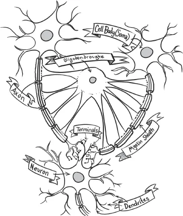
FIGURE 1.1. Brain cells include communicators (neurons) and supporters (glia). Both have a cell body (soma) that has structures to keep the cell alive. Neurons communicate by sending electrical impulses (action potentials) down a wire-like structure (axon) that forms a connection (synapse) that almost touches the branches (dendrites) of the next neuron. Communicating molecules (neurotransmitters) are released into this space, binding to receptors on the next cell’s dendrites. Glia insulate axons with a fatty coating (myelin sheath) and help to clean up nearby molecules and neurotransmitters.
Each neuron communicates with other neurons by building up an electrical charge that causes a cell’s axon to shoot chemicals across the small gap between itself and a downstream cell’s dendrites. This gap is called the synaptic cleft. These chemicals (known as neurotransmitters and neuromodulators) change the voltage of the downstream cell, making it more or less likely to fire its own action potential. This transmission process is the fundamental computation of the brain: one cell decides to fire (or not) based on the signals that the cells connected to it send (or don’t). We’ll discuss this a bit more in the next chapter.
But what about those other cells that we mentioned, the glia? Well, for a long time most neuroscientists thought that they were sort of like the support staff for neurons. They clean up the messes that happen when neurons shoot those neurotransmitters all over the place. They also help keep neurons healthy and foster communication between cells. While this support staff model of glia seems accurate as far as it goes, it is becoming increasingly apparent that glia are so much more than that. Each year, more studies come out showing that glia are also doing a little bit of computing on their own. However, what this computing is and how it relates to behavior is still a big mystery.
But how does all of this make the brain work?3
We’ve known for some time that the brain is a massive interconnected network. Of course, early estimates of how massive this network is were a bit overstated. Take, for example, the headline of an article that ran in the New York Times on June 25, 1933, “Brain Phone Lines Counted as 1 Plus 15 Million Zeros: Scientists Told of Figures So Stupendous That Astronomy Fades in Comparison.” Assuming what we know about the size of neurons and their axons, this would require that your brain take up an area slightly larger than the solar system. But while this number was just a little bit inflated, there are in fact a lot of neurons: somewhere between 80 and 100 billion cells with anywhere from a hundred to tens of thousands of connections from each. So basically, the brain functions as a massively connected computer network, one with trillions (with a “t”) of connected parts.
To put this into perspective, based on reports by the computer networking company Cisco, as of 2013 there were about 10 billion active connections on the entire internet.4 The entire internet will not even reach 50 billion connections until the year 2020. This means that your brain is almost 10 times more connected than the entire internet is right now.
However, if you take a step back and look at a brain without a microscope, the first thing you notice is that it looks very wrinkly. The tissue folds over itself like the face of a Shar-Pei dog. That’s because there’s barely enough room in our skulls to fit all of those cells. So the tissue gets squished in there as compactly as possible. The mountains of the folds are called gyri (or gyrus if you’re talking about just one) and the valleys are called sulci (or sulcus for just one)....
Table of contents
- Cover Page
- Title Page
- Copyright Page
- Contents
- List of Figures
- Prelude: Sacrifices Not Made in Vain
- Introduction
- Chapter 1: Gray’s (Undead) Anatomy
- Chapter 2: Do Zombies Dream of Undead Sheep?
- Chapter 3: The Neural Correlates of Lumbering
- Chapter 4: Hungry, Angry, and Stupid is No Way to Go Through Unlife
- Chapter 5: There’s No Crying in the Zombie Apocalypse!
- Chapter 6: Tongue-tied and Twisted
- Chapter 7: Disengagement Deficit of the Dead
- Chapter 8: Whose Undead Face Is This, Anyway?
- Chapter 9: How Am I Not Myself?
- Chapter 10: Eternal Sunshine of the Undead Mind
- Chapter 11: Fighting the Zombie Apocalypse … With Science!
- Acknowledgments
- Glossary
- Index
Frequently asked questions
Yes, you can cancel anytime from the Subscription tab in your account settings on the Perlego website. Your subscription will stay active until the end of your current billing period. Learn how to cancel your subscription
No, books cannot be downloaded as external files, such as PDFs, for use outside of Perlego. However, you can download books within the Perlego app for offline reading on mobile or tablet. Learn how to download books offline
Perlego offers two plans: Essential and Complete
- Essential is ideal for learners and professionals who enjoy exploring a wide range of subjects. Access the Essential Library with 800,000+ trusted titles and best-sellers across business, personal growth, and the humanities. Includes unlimited reading time and Standard Read Aloud voice.
- Complete: Perfect for advanced learners and researchers needing full, unrestricted access. Unlock 1.5M+ books across hundreds of subjects, including academic and specialized titles. The Complete Plan also includes advanced features like Premium Read Aloud and Research Assistant.
We are an online textbook subscription service, where you can get access to an entire online library for less than the price of a single book per month. With over 1.5 million books across 990+ topics, we’ve got you covered! Learn about our mission
Look out for the read-aloud symbol on your next book to see if you can listen to it. The read-aloud tool reads text aloud for you, highlighting the text as it is being read. You can pause it, speed it up and slow it down. Learn more about Read Aloud
Yes! You can use the Perlego app on both iOS and Android devices to read anytime, anywhere — even offline. Perfect for commutes or when you’re on the go.
Please note we cannot support devices running on iOS 13 and Android 7 or earlier. Learn more about using the app
Please note we cannot support devices running on iOS 13 and Android 7 or earlier. Learn more about using the app
Yes, you can access Do Zombies Dream of Undead Sheep? by Timothy Verstynen,Bradley Voytek in PDF and/or ePUB format, as well as other popular books in Psychology & Cognitive Neuroscience & Neuropsychology. We have over 1.5 million books available in our catalogue for you to explore.