The potential impact of anthropogenic pollutants such as agrochemicals on the environment is of global concern. Increasing use of certain compounds can result in contamination of food, water and atmospheric systems and in order to combat this pollution it is important to be able to accurately monitor the short and long term effects.
This book describes the latest non-traditional terrestrial species models used as indicators of the toxic effects of environmental pollutants. The book enables understanding of the effects of pollutants in non-target species, and therefore enables analysis of the effects on ecosystems.
This book will be of interest to anyone interested in developing new biomarker species with high degrees of ecological relevance. It will serve as a useful resource for regulatory and research toxicologists, particularly those interested in soil screening and the effects of pollutants on wildlife and insects and their use as biological indicators.
- 340 pages
- English
- ePUB (mobile friendly)
- Available on iOS & Android
eBook - ePub
About this book
Trusted by 375,005 students
Access to over 1.5 million titles for a fair monthly price.
Study more efficiently using our study tools.
Information
Section II: Terrestrial Vertebrates as Experimental Models
CHAPTER 6
Use of Melanin-pigmented Cells as a New Tool to Evaluate Effects of Agrochemicals and Other Emerging Contaminants in Brazilian Anurans
a Department of Biology, São Paulo State University (UNESP), São José do Rio Preto, São Paulo, Brazil;
b Goiano Federal Institute (IF Goiano), campus Rio Verde, Goiás, Brazil
*E-mail: [email protected]
6.1Color in Animals
Specialized cells for storing pigments, chromatophores, are found in invertebrate and vertebrate ectotherms. These cells show various cytoplasmic projections and are originated from the embryonic neural tube; then, they migrate to the skin and are distributed on the epidermis and dermis.1
There are various types of pigments found in chromatophores. In vertebrates, at least five of them have been described. Melanophores, which are black or brown, contain melanin in granules. Erythrophores, with their reddish color, contain pteridine; xanthophores also have pteridine, besides carotenoids located in vesicles, which give them a yellow color. Iridophores have a metallic color, owing to the presence of purines deposited in reflecting crystals. These four types of pigmented cells are found in fish, amphibians, and reptiles. Leucophores, on the other hand, contain purine granules, which give them a white color, and are found only in fish.1,2 Our main analyses and observations are based on a very common type of extracutaneous melanophore, called visceral melanocyte, which is discussed further on (Figure 6.1).
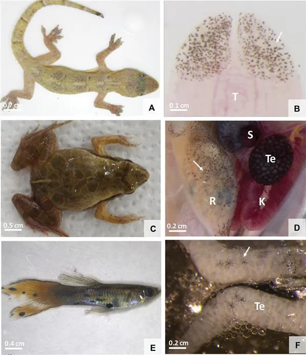
Figure 6.1External and visceral colors in ectothermic animals (A, B: Reptile, Hemidactylus mabouia; C, D: Anuro, Physalaemus cuvieri, E, F: Fish, Poecilia reticulata and Knodus moenkhausii). Different color patterns given by chromatophores (A, C, E) besides the presence of visceral melanocytes (arrows) with a dendritic aspect, which give the organs a dark color. A: H. mabouia natural color. B: Melanocytes present (arrow) in the ventral surface of the tongue tip. C: P. cuvieri color, showing the coloration pattern. D: Cells containing melanin (arrows) in organs. E: Cutaneous color of P. reticulata. F: Presence of pigmented cells in K. moenkhausii testes. K: Kidney. R: Rectum. S: Spleen. T: Tongue. Te: Testes.
Even though the term melanophore is widely used by several authors,3 in order to simplify the nomenclature and to recognize the increasing evidence of a conservation genetics of the melanocytes biology, the term melanocytes was proposed to be applied to all those cells. However, since the term melanophore has been used by different authors and it is also correct, it may still be used.
An advantage in introducing this additional category of melanocytes (melanophores) is to create a more precise classification of the cells’ behavior, but it has not been incorporated yet, such as by other subtypes of melanin-secreting melanocytes in mammals, which have not been treated similarly. We opted to use the term melanocyte for cells that contain melanin in the internal organs of ectothermic animals and melanophore for melanin-containing cells in skin.
Cutaneous chromatophores are usually found on the dermis, where erythrophores are the most superficial and melanophores are the deepest ones (Figure 6.2). The arrangement of these pigment cells over the different skin layers is related to the type of pigment they contain and which wavelengths they reflect and absorb, thus providing vertebrates with different colors.2
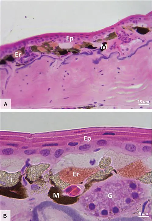
Figure 6.2Histology of Physalaemus nattereri skin. A: General view of the skin section showing an erythrophores layer (Er), found under the epidermis (Ep), followed by a deeper melanophores layer (M). B: Chromatophores found in the skin: erythrophores (Er), iridophores (I) and melanophores (M), with their different colors. G: exocrine gland.
In fish, some chromatophores respond to changes in the environment, which might interfere with the animals’ capacity to interact with their environment (e.g., camouflage, cryptic colorations). Recently, authors4 have shown that in fish (Oreochromis mossambicus) melanophores respond to lead nitrate, phenol, and hexachlorocyclohexane. In another fish, Channa punctatus, upon exposure to arsenic, the dispersion of melanophores was decreased at initial exposure times (i.e., up to 60 days). As a result, the animal's color becomes lighter; however, after 90 days, dispersion increases, and the animal's color becomes darker. This shows that the fish develop physiological response mechanisms to the exposure to arsenic.5
According to this approach, in ectothermic animals, the chromatophores that synthesize dark pigments, called melanocytes, are not only limited to the dermis, but frequently occur in connective tissue and in internal organs, such as the liver, kidneys, heart, thymus, gonad, besides blood vessels, peritoneum and meninges.2,6
6.2Internal Melanin-pigmented Cells
Both fish and amphibians have pigmented cells in their organs and membranes, called internal melanocytes.7–10 These cells are originated from the ectothermic neural crest;11 they contain large quantities of melanin6 and are similar to dermal melanocytes.12 They also have various dendritic extensions, which can even be observed without a microscope.13
There are other, different, pigmented cells called melanomacrophages, which are present in hematopoietic organs (e.g., liver and spleen) and have phagocytic activity similar to macrophages.14,15 These cells have substances from cellular catabolism in the cytoplasm, such as hemosiderin and lipofuscin.14 Melanomacrophages are derived from hematopoietic stem cells and are round in shape.11,16
In common, and our main interest in this chapter, is the melanin present both in melanocytes and melanomacrophages; it is a complex polymer synthesized endogenously in vertebrates and invertebrates17 that absorbs and neutralizes free radicals and other potentially toxic agents resulting from catabolism.14 Furthermore, melanin plays an immune role in ectothermic animals, owing to the action of hydrogen peroxidases and their quinone precursors, which act as bactericides through the increase in enzymatic activity, even though this is restricted to low temperatures.18 Another function attributed to melanin is related to photoreception and thermoregulation in ectothermic animals, besides photoprotection, acting as a photosensibilizer in cells exposed to radiation with enough energy to cause genetic damage.19
In this context, for example, for certain anuran amphibians, both testicular melanocytes and hepatic melanomacrophages respond to the administration of lipopolysaccharide (LPS) from Escherichia coli with an increase in the pigmented area, which suggests that these cells have tissue protection and bactericide functions.13 The testicular pigmentation occurs in some species and, from the evolutionary and phylogenetic relationship points of view, it also varies among anuran families.20 This internal pigmentation is a characteristic conserved throughout phylogeny in all organs.21
Visceral pigmentation in fish, in turn, from a functional point of view, might be related to the accumulation of residual melanin; it could also play a role in the innate immune system, have antioxidant functions, and protect the tissue against damages in the DNA.20 However, visceral pigmentation functions in fish and anurans have been little studied and it is necessary to deepen this investigation in order to test how such pigmentation responds to external stimuli, since both transparent and non-transparent animals show internal organ pigmentation.20
6.3Environmental Contamination and Its Effects on Visceral Pigmentation
Fish and amphibians live in aquatic environments, where contaminants are frequently found. Thus, these animals represent good indicators of environmental quality.22...
Table of contents
- Cover
- Title
- Copyright
- Preface
- Contents
- Section I: Terrestrial Invertebrates as Experimental Models
- Section II: Terrestrial Vertebrates as Experimental Models
- Subject Index
Frequently asked questions
Yes, you can cancel anytime from the Subscription tab in your account settings on the Perlego website. Your subscription will stay active until the end of your current billing period. Learn how to cancel your subscription
No, books cannot be downloaded as external files, such as PDFs, for use outside of Perlego. However, you can download books within the Perlego app for offline reading on mobile or tablet. Learn how to download books offline
Perlego offers two plans: Essential and Complete
- Essential is ideal for learners and professionals who enjoy exploring a wide range of subjects. Access the Essential Library with 800,000+ trusted titles and best-sellers across business, personal growth, and the humanities. Includes unlimited reading time and Standard Read Aloud voice.
- Complete: Perfect for advanced learners and researchers needing full, unrestricted access. Unlock 1.5M+ books across hundreds of subjects, including academic and specialized titles. The Complete Plan also includes advanced features like Premium Read Aloud and Research Assistant.
We are an online textbook subscription service, where you can get access to an entire online library for less than the price of a single book per month. With over 1.5 million books across 990+ topics, we’ve got you covered! Learn about our mission
Look out for the read-aloud symbol on your next book to see if you can listen to it. The read-aloud tool reads text aloud for you, highlighting the text as it is being read. You can pause it, speed it up and slow it down. Learn more about Read Aloud
Yes! You can use the Perlego app on both iOS and Android devices to read anytime, anywhere — even offline. Perfect for commutes or when you’re on the go.
Please note we cannot support devices running on iOS 13 and Android 7 or earlier. Learn more about using the app
Please note we cannot support devices running on iOS 13 and Android 7 or earlier. Learn more about using the app
Yes, you can access Ecotoxicology and Genotoxicology by Marcelo L Larramendy in PDF and/or ePUB format, as well as other popular books in Medicine & Toxicology. We have over 1.5 million books available in our catalogue for you to explore.