The ultra-bright femtosecond X-ray pulses provided by X-ray free electron lasers (XFELs) open up opportunities to study the structure and dynamics of a wide variety of systems beyond what is possible with synchrotron sources. This book introduces the principles and properties of currently operating and future XFELs, before outlining applications in materials science, chemistry and biology. Edited by pioneers in this exciting field, and featuring contributions from leading researchers, this book is ideal for researchers working with XFELs, synchrotron radiation, ultrafast and femtosecond crystallography and femtosecond spectroscopy.
eBook - ePub
X-Ray Free Electron Lasers
Applications in Materials, Chemistry and Biology
- 463 pages
- English
- ePUB (mobile friendly)
- Available on iOS & Android
eBook - ePub
X-Ray Free Electron Lasers
Applications in Materials, Chemistry and Biology
About this book
Trusted by 375,005 students
Access to over 1.5 million titles for a fair monthly price.
Study more efficiently using our study tools.
Information
Section IV
Photochemistry in Materials
CHAPTER 8
Gas Phase Photochemistry Probed by Free Electron Lasers
THOMAS J. A. WOLF*a AND MARKUS GÜHR*a,b
aSLAC National Accelerator Laboratory, Stanford PULSE Institute, 2575 Sand Hill Road, Menlo Park, 94025-7015, USA
bPotsdam University, Institute for Physics and Astronomy, Karl-Liebknecht-Straße 24/25, 14476 Potsdam-Golm, Germany
*E-mail: [email protected], [email protected]
8.1 Introduction
The most important interactions between molecules in nature and sunlight in the visible and ultraviolet (UV) spectral regime, including the initial steps in light harvesting,1 human vision2 and DNA photoprotection,3 involve ultrafast excited state dynamics on the femtosecond and picosecond timescale. Since the evolution of picosecond and femtosecond laser systems, a whole research field has evolved between chemistry and physics, investigating excited state dynamics in a plethora of samples ranging from small model systems in the gas phase to biomolecules in their natural environment.
The time resolved investigation of fundamental processes in isolated gas phase molecules can lead to intimate understanding of the underlying electronic and geometric mechanisms of ultrafast dynamics. The complexity of the investigated system and of the methods can be well matched to the complexity of the questions to be answered. Such investigations can benefit in particular from support by high-level quantum chemical calculations on isolated molecules.4–26 The possibility to simulate experimental observables thereby allows for fruitful joint experimental and theoretical studies.11,27–29
The choice of the wavelength used to trigger excited state dynamics is usually determined by the absorption spectrum of the molecule. The wavelength used to probe the dynamics is considerably more flexible. For isolated targets, the most common observable is the yield and kinetic energy of charged particles. Thus, the molecule needs to be ionized by the probe laser pulse. This can be achieved through various schemes, most conveniently by multiphoton ionization using strong infrared (IR) pulses available from Ti:Sapph lasers.30,31 For most molecules, UV pulses allow the ionization of excited states,5,32–39 although the probe range might be limited by decreased Franck–Condon factors and increasing ionization potentials due to molecular relaxation processes.40,41 This problem can be overcome using probe pulses with much higher energy in the vacuum UV range, as the first examples show.42–46
Valence photoelectron or photoion spectroscopy has delivered deep insight into many ultrafast molecular processes. Ultrafast spectroscopy involving the 1s core electrons during the probe process will, however, deliver complementary information. For organic molecules, the K-edges of carbon, nitrogen and oxygen are most important in this context. The 1s binding energy differs by more than 100 eV from one element to the next, allowing one to probe molecular processes from an element-specific point of view. Core electrons are bound extremely tightly around the specific nucleus and are highly symmetric. Thus, a probe transition involving the 1s levels is highly local and site specific and the selection rules depend mostly on the final state, which is either an unoccupied orbital or the photoionization continuum.
A rich amount of literature on core level transitions of isolated molecules was developed at synchrotron sources.47–59 Now that ultrafast and strong soft X-ray (SXR) sources are available due to the advancement of X-ray free electron lasers (XFELs),60–62 the spectroscopic knowledge can be used for element- and site-specific spectroscopy of ultrafast photoexcited molecular processes.
This chapter reviews essential methods for probing dynamics of isolated molecules. We give specific examples for X-ray induced Auger and photoion probing, and discuss promising future absorption and photoelectron probing.
8.2 Different Ways of Probing the Molecular Dynamics: Direct vs. Indirect
We divide the X-ray probe methods that are available for isolated molecules into two classes, as shown in Figure 8.1. For the direct methods, which are photoabsorption and electron photoemission, we observe the final state of the molecule in the presence of an X-ray induced 1s core hole. The 1s electron can be resonantly transferred into an unoccupied or partially valence state, as in near edge X-ray absorption fine structure (NEXAFS) spectroscopy.63 The NEXAFS spectrum is directly related to the electronic structure and this information will be available in a time resolved fashion using short X-ray resonant probe pulses. Alternatively, for photon energies above the core level ionization potential, the photoelectron kinetic energy can be determined, as in photoemission spectroscopy. Studies on electronvolt scale splitting show that the exact kinetic energy of the photoelectron depends on the electronegativity of the nearest environment of the particular 1s level under investigation.48,64,65
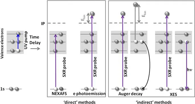
Figure 8.1 UV pump–X-ray probe scheme showing several options for detecting the probe step. In the direct methods, either resonant absorption to partially filled or unfilled valence states or the continuum are detected. The indirect methods monitor decay products of the core hole decay, which can be photons or Auger electrons, as well as the photoions resulting from the molecular Auger decay.
In time resolved measurements, these aspects can be used to follow nuclear, as well as electronic dynamics after photoexcitation. The direct methods rely on a spectrally stable X-ray source. Jitter in spectral shape and position translates to large line broadening in the absorption, as well as in photoelectron spectra. At XFEL sources starting from noise via self-amplified spontaneous emission (SASE), these methods are still challenging and conditions are, by far, not ideal for these types of spectroscopy.66 The first experiments at XFELs therefore used so called “indirect” X-ray methods, where observables do not explicitly depend on photon energy, as shown in Figure 8.1.
For indirect methods, one detects the decay products of the core hole relaxation. Thus, the final state of the probe process does not contain a 1s core hole any more. A core hole has a limited lifetime, which is typically a few femtoseconds in the SXR domain. The major core hole decay process in the photon energy range from the carbon to the oxygen K-edge (from 280 to 540 eV) is Auger decay resulting in the emission of an Auger electron with a few hundred electronvolts of kinetic energy. X-Ray photoemission is far more rare (below 0.1% compared to Auger), and thus has limited use for very dilute gas phase targets. That might, however, change with the next generation of XFELs with MHz repetition rates and higher averaged flux coming online in the close future.67 For photoionization above the continuum, the Auger electron energy, as well as the X-ray photoemission wavelength do not depend on the energy of the ionizing photon. This makes the methods ideal for spectrally fluctuating SASE sources. The theoretical description for molecular Auger processes is far more complex than the direct processes, and highly congested spectra already for the ground state59 indicate that details of excited state spectra are hard to simulate for open shell excited states. The non-resonant Auger decay leaves a minimum of two positive charges on the molecule, which leads to molecular fragmentation. The mass and kinetic energy of these fragments can be analyzed and related to the photoexcited molecular dynamics, as discussed later.
8.2.1 Indirect Methods
In this section, we present three experiments performed in the first years of the Linac Coherent Light Source (LCLS). The first experiment uses the Auger decay as an indication of molecular nuclear relaxation and the following internal conversion of the UV excited nucleobase thymine. The other two examples use the detection of Auger induced fragmentation to investigate ultrafast photoinduced ring opening and bond dissociation reactions.
8.2.1.1 Auger Electron Spectroscopy
Nucleobases possess a very strong UV absorption; however, they show remarkable photostability. It is argued that this was an import...
Table of contents
- Cover
- Title
- Copyright
- Preface
- Quote
- Contents
- Section I: Properties of XFELs
- Section II: Biological Structure Determination
- Section III: Photochemistry in Biological Systems
- Section IV: Photochemistry in Materials
- Section V: Sample Delivery Methods
- Section VI: New Directions
- Subject Index
Frequently asked questions
Yes, you can cancel anytime from the Subscription tab in your account settings on the Perlego website. Your subscription will stay active until the end of your current billing period. Learn how to cancel your subscription
No, books cannot be downloaded as external files, such as PDFs, for use outside of Perlego. However, you can download books within the Perlego app for offline reading on mobile or tablet. Learn how to download books offline
Perlego offers two plans: Essential and Complete
- Essential is ideal for learners and professionals who enjoy exploring a wide range of subjects. Access the Essential Library with 800,000+ trusted titles and best-sellers across business, personal growth, and the humanities. Includes unlimited reading time and Standard Read Aloud voice.
- Complete: Perfect for advanced learners and researchers needing full, unrestricted access. Unlock 1.5M+ books across hundreds of subjects, including academic and specialized titles. The Complete Plan also includes advanced features like Premium Read Aloud and Research Assistant.
We are an online textbook subscription service, where you can get access to an entire online library for less than the price of a single book per month. With over 1.5 million books across 990+ topics, we’ve got you covered! Learn about our mission
Look out for the read-aloud symbol on your next book to see if you can listen to it. The read-aloud tool reads text aloud for you, highlighting the text as it is being read. You can pause it, speed it up and slow it down. Learn more about Read Aloud
Yes! You can use the Perlego app on both iOS and Android devices to read anytime, anywhere — even offline. Perfect for commutes or when you’re on the go.
Please note we cannot support devices running on iOS 13 and Android 7 or earlier. Learn more about using the app
Please note we cannot support devices running on iOS 13 and Android 7 or earlier. Learn more about using the app
Yes, you can access X-Ray Free Electron Lasers by Uwe Bergmann, Vittal Yachandra, Junko Yano in PDF and/or ePUB format, as well as other popular books in Physical Sciences & Chemistry. We have over 1.5 million books available in our catalogue for you to explore.