Order the Set Medical Physics and save almost 25€.
Medical Physics covers the applied branch of physics concerned with the application of concepts and methods of physics to diagnostics and therapeutics of human diseases. This second volume in a series of two complements the imaging modalities presented in the first volume by those methods, which use ionizing radiation. The first chapters in part A on Radiography provide a solid background on radiation sources, interaction of radiation with matter, and dosimetry for the safe handling of radiation before introducing x-ray radiography, scintigraphy, SPECT and PET.
The second part B on Radiotherapy starts from basic information on the life cycle of cells, radiation response of healthy and tumorous cells. In subsequent chapters the main methods of radiation treatment are presented, in particular x-ray radiotherapy, proton and neutron radiation therapy, and brachytherapy. The last part C, Diagnostics and Therapeutics beyond Radiology, covers laser applications, multifunctional nanoparticles and prosthetics.
The present volume
- introduces the physical background on ionizing radiation, the biological effectiveness of radiation, as well as radiation based methods for diagnostics and therapeutics.
- covers the second part of the entire field of medical physics, including imaging methods with the use of ionizing radiation; radiation therapy with photons, protons, and neutrons; laser methods, nanomedicine and prosthetics.
- provides an introduction for Bachelor students to the main concepts of Medical Physics during their fi rst semesters guiding them to further specialized and advanced literature.
- contains many questions & answers related to the content of each chapter.
- is also available as a set together with Volume 1.
Contents
Part A: Radiography
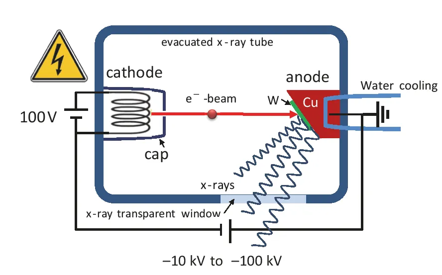
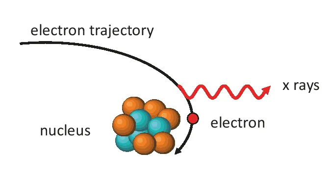
X-ray generation
Nuclei and isotopes
Interaction of radiation with matter
Radiation detection and protection
X-ray radiography
Scintigraphy
Positron emission tomography
Part B: Radiotherapy
Cell cycle and cancer
X-ray radiotherapy
Charged particle radiotherapy
Neutron radiotherapy
Brachytherapy
Part C: Diagnostics and therapeutics beyond radiology
Laser applications in medicine
Nanoparticles for nanomedical applications
Prosthetics