![]()
HEAD AND NECK
![]()
12
Congenital Neck Masses
Andrew R. Scott and Michael J. Cunningham
INTRODUCTION
Most neck masses in children are benign lesions of inflammatory origin that either respond to medical therapy or resolve spontaneously. Excisional biopsy is usually indicated for diagnostic or cosmetic purposes in the case of recurrent or persistent neck masses. Therapeutic surgical intervention is warranted for lesions that cause airway symptoms, dysphagia or present with other worrisome features. This chapter will focus on pediatric neck masses of congenital etiology.
The term congenital literally means present at birth, however, many of the lesions described in this chapter do not become apparent until later in childhood and occasionally in adulthood. A working knowledge of the embryonic events leading to the formation of these lesions improves understanding of their pathophysiology and enhances their effective surgical removal.
The following is a review of the presentation, work-up and management of the more common congenital neck masses organized according to the characteristic age of presentation during childhood. Congenital vascular anomalies also arise in the head and neck region in children; details regarding such infantile hemangiomas and vascular malformations of lymphatic and venous origin can be found elsewhere in this book.
NECK MASSES PRESENTING IN THE FETAL OR NEONATAL PERIOD
Teratomas and Congenital High Upper Airway Obstruction (CHAOS)
Teratomas refer to tumors that are composed of cells from all three germ cell layers — ectoderm, mesoderm, and endoderm — in various stages of maturity and are comprised of tissue that is foreign to the site of origin. Teratomas most commonly occur in the sacrococcygeal and cervicofacial regions. Cervical teratomas are characteristically benign lesions. They are frequently diagnosed in utero with a 20% to 50% incidence of maternal polyhydramnios thought to be related to extrinsic esophageal obstruction; additionally alpha fetal protein levels may be elevated.1
The fetal presentation of a teratoma may be associated with congenital high upper airway obstruction syndrome (CHAOS), a rare in utero condition characterized by complete or near complete obstruction of the upper airway. Potential etiologies of CHAOS include laryngeal atresia or stenosis, tracheal atresia or stenosis, or extrinsic upper airway compression due to a large mass in the neck or upper aerodigestive tract. While there are case reports of foregut duplication cysts and lymphatic vascular malformations causing such upper airway obstruction, teratomas represent the vast majority of congenital tumors associated with CHAOS.2
The diagnosis of CHAOS is usually made during prenatal ultra-sound. Characteristic findings include polyhydramnios, lung expansion, inversion of the diaphragm and cardiac compression. When due to extrinsic compression, the large cervical or upper aerodigestive tract mass is typically identifiable.
Further work-up necessitates the assistance of a maternal-fetal medicine specialist or perinatologist in addition to the potential consultative services of a medical geneticist, a pediatric surgeon, and a pediatric otolaryngologist. Fetal echocardiogram and amniocentesis for karyotype may be obtained. Magnetic resonance imaging allows for excellent soft tissue definition without the potential morbidity associated with radiation to the developing fetus. Such high resolution fetal MRI of the head and neck can prove particularly useful in cases of extrinsic airway compression from large neck masses that may need to be addressed at birth.
Ultimately an airway must be established at the time of delivery. This is typically accomplished via a so-called ex utero intrapartum treatment (EXIT) procedure. Such an EXIT procedure is often performed after 30 weeks gestation if possible, balancing early intervention with the survival benefit afforded by maximizing fetal size and lung maturity. Other factors such as fetal hydrops or congestive heart failure may necessitate earlier delivery. During the EXIT procedure, the mother is maintained under general anesthesia while the head and neck of the fetus is partially delivered through an incision in the uterus. The placenta remains intact, and fetal perfusion is maintained through the umbilical circulation, a scenario analogous to extracorporeal membrane oxygenation (ECMO). This technique allows for up to 90 minutes of time in which the fetal airway can be secured either through endoscopic-assisted intubation or by surgical means. Additionally there are reports of subtotal resection of cervical teratomas during the EXIT procedure in order to facilitate establishment of an airway.3
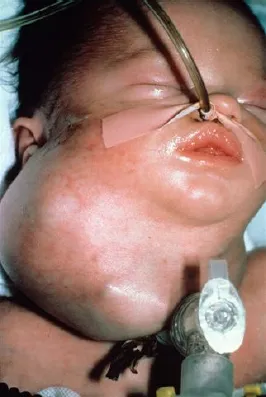
Those cervical teratomas not identified prenatally are typically evident at the initial neonatal assessment. Examination usually reveals a firm, anterolateral neck mass which may display nodularity and focal areas of discoloration (Fig. 1). These masses are often quite large with secondary tracheal compression or deviation.
Radiographic evaluation of a cervical teratoma with computed tomography (CT) or magnetic resonance imaging (MRI) allows for high resolution evaluation of the mass itself and definition of the tumor’s relationship to adjacent structures. These imaging modalities typically reveal a well-encapsulated, heterogeneous lesion with both solid and cystic components; there may be speckled calcifications on CT imaging.
Surgical resection is the treatment of choice for cervical teratomas. Complete excision, if feasible, is ideal. These masses are usually poorly vascularized. Operative morbidity includes potential paresis or paralysis of the vagus and recurrent laryngeal nerves on the affected side. The recurrent laryngeal nerve is at particular risk due to the fact that these tumors are often intimately related to the thyroid gland. Near total thyroidectomy may be required to achieve adequate tumor margins. Postoperatively these patients should be followed with regular examinations to assess for recurrence and to monitor for hypothyroidism.1
Fig. 1. Infant with a cervical teratoma requiring tracheotomy for airway securement at birth.
Fibromatosis Colli
Pseudotumor of infancy (POI) is a unilateral contracture of the sternocleidomastoid (SCM) muscle that may be evident at birth or become manifest within the first six weeks of life. The SCM contracture causes a characteristic posture in which the chin is rotated away from the lesion and the occiput is rotated towards the affected side, hence the term congenital muscular torticollis.
On physical examination a firm, non-tender mass is palpable within the body of the contracted SCM muscle. This mass, known as fibromatosis colli, occurs when muscle tissue is replaced by fibrosis. The exact cause is unknown but several theories exist. The intrauterine theory postulates that compressive forces lead to abnormal fetal positioning of the neck causing fibrosis and contracture of the SCM. The traumatic theory poses that there is damage to the SCM during a difficult delivery that leads to intramuscular hematoma and scarring. Other etiologies such as compartment syndrome, vascular occlusion leading to ischemia and infarction, and even a genetic influence have been postulated. Although the clinical presentation is typically straightforward, ultrasound is the diagnostic study of choice to confirm the mass in question is truly intramuscular.4 A solid mass within the sternocleidomastoid muscle is consistent with fibromatosis colli. Surgical biopsy is necessary only when the ultra-sonographic evaluation alternatively documents an extramuscular lesion raising concern of a more worrisome diagnosis such as a peripheral nerve sheath tumor or a cervical neuroblastoma.
The natural history of fibromatosis colli is usually one of spontaneous regression. Pseudotumor of infancy and congenital muscular torticollis are therefore best managed expectantly. Physical therapy for passive range of motion exercises may enhance recovery. Surgery is indicated only in those infrequent cases in which the torticollis fails to resolve after 12 to 18 months.5 Facial asymmetry and plagiocephaly may become apparent in these children over time. Surgical intervention is in the form of a sternocleidomastoid tenotomy to release the contracture.
NECK MASSES PRESENTING IN INFANCY AND CHILDHOOD
Midline Neck Masses
Thyroid gland anomalies
During the third week of gestation, the thyroid gland develops from a diverticulum arising from the floor of the pharynx and becomes bilobed as it descends into the anterior neck. Throughout its migration, the primitive gland remains attached to its origin within the pharynx by a hollow canal, the thyroglossal duct, which ultimately involutes by weeks 5 to 8 of gestation. The pharyngeal site of attachmen...