![]()
CHAPTER 1
AN INTRODUCTION TO COMPUTER VISION IN
MEDICAL IMAGING
There has been much progress in computer vision and pattern recognition in the last two decades, and there has also much progress in recent years in medical imaging technology. Although images in digital form can easily be processed by basic image processing techniques, effective use of computer vision can provide much useful information for diagnosis and treatment. It has been a challenge to use computer vision in medical imaging because of complexity in dealing with medical images. In this chapter a brief introduction of the subject is presented by addressing the issues involved and then focusing on the active contour model for medical imaging.
1. Introduction
There has been enormous progress in medical imaging techniques and modalities in the last decade or so. For example ultrasound has found its use in many areas previously using x-ray or other techniques. Accompanied with the progress is the greatly increased use of computer vision techniques in medical imaging. In computer vision (see e.g. [1][2]) we talk about the low-level processing which involves basic image processing operations like noise filtering, contrast enhancement and image sharpening, the mid-level processing which involves image segmentation and pattern recognition as well as 3D reconstruction and the high-level processing which involves ‘making sense’ of an ensemble of recognized objects and performing the cognitive functions at the far end of the processing sequence. Medical imaging refers to the techniques and processes used to create images of the human body for clinical purposes, or procedures seeking to reveal, diagnose or examine disease or studying normal anatomy and physiology [3]. Medical imaging evolved from the discovery of x-rays to the newest magnetic resonance image (MRI). The most commonly used techniques these days are x-ray, computer tomography (CT), ultrasound, MRI and positron emission tomography (PET). The emphasis of medical imaging is to help doctors or other trained personal to provide better diagnosis and treatment and thus the low level and mid-level computer vision is particularly important in the medical area. It is evident that medical imaging has significant impact on medicine and computer vision making use of enormous computing power has enormous impact on medical imaging. The following sections will briefly discuss the nature of some of these medical images and the technology behind them. The remaining chapters of the book cover important aspects of computer vision in medical imaging written by leading experts in the field.
2. Some Medical Imaging Methods
2.1. X-ray
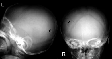
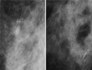
X-ray is the first and oldest medical technique available to doctors for the visualization of the body without surgery. X-rays were first discovered by Wilhelm Rontgen in 1895. They penetrate most biological tissues with little attenuation and thus provide a comparatively simple means to produce shadow or projection, images of human body. However X-rays have ionizing effects on the body and hence should not be repeatedly used. The X-ray imaging system involves having a film or screen containing a radiation-sensitive material exposed to the x-rays transmitted through the region of the body. The developed film or excited phosphorous screen exhibits a geometric pattern produced by the structures in the beam path [4]. However X-ray imaging is limited as the signal can be reduced due to the scattering of a large percentage of radiation from the body and much detail is lost in the radiographic process with the superposition of 3D structural information onto a 2D surface. Fig. 1 is the X-ray image of the bones. X-ray system has now been greatly improved. Its use for digital mammogram is particularly important (see e.g. Fig. 2, based on our 1996 data base [14]).
Fig. 1. X-ray image of a skull with a fracture in the right parietal bone.
Fig. 2. Mammograms of a patient.
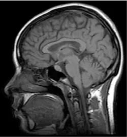
Fig. 3. MRI image of the human head.
2.2. Magnetic Resonance Image (MRI)
Magnetic Resonance Image was developed in the early 1970s and has become versatile and clinically useful diagnostic imaging modality [4,5]. MRI is a noninvasive imaging technology that does not use ionizing radiation and provides much more contrast between different soft tissues of the body than computed tomography (CT). It is based on perturbing magnetic fields with radio waves. In MRI, hydrogen nuclei (protons) are imaged due to their strong magnetic moment and prevalence in the soft tissue of the body. The magnetic field is produced by passing an electric current through wire coils in most MRI units. Other coils, located in the machine and in some cases, placed around the part of the body being imaged, send and receive radio waves, producing signals that are detected by the coils. A computer then processes the signals and generates a series of images each of which shows a thin slice of the body. The images can then be studied from different angles by the interpreting physician. Fig. 3 shows the MRI image of the human head. MRI studies brain or body anatomy. More recently, functional MRI has been particularly useful to study brain physiologic function.
2.3. Intravascular Ultrasound (IVUS)
Intravascular Ultrasound (or IVUS) allows us to see the coronary artery from the inside-out. This unique picture, Fig. 4, generated in real time, yields information that is not possible with routine imaging methods or even non-invasive Multislice CT scans. A growing number of cardiologists think that new information yielded by IVUS can make a significant difference in how patient is treated and can provide more accurate information which will reduce complications and incidence of heart diseases. Intravascular ultrasound (IVUS) is a catheter-based technique which provides high-resolution images allowing precise tomographic assessment of lumen area. IVUS uses high-frequency sound waves called ultrasound that can provide a moving picture of your heart. These pictures come from inside the heart rather than through the chest wall. The sound waves are sent with a device called a transducer. The transducer is attached to the end of a catheter, which is threaded through an artery and into your heart. The sound waves bounce off of the walls of the artery and return to the transducer as echoes. The echoes are converted into images on a television monitor to produce a picture of your coronary arteries and other vessels in your body.
Fig. 4. Typical IVUS image.
In the above IVUS image, the lumen is typically a dark echo-free area adjacent to the imaging catheter and the coronary artery vessel wall mainly appears as three layers: intima, media, and adventitia. As the two inner layers are of principal concern in clinical research, segmentation of IVUS images is a must to isolate the intima-media and lumen boundaries which provides important information about the degree of vessel obstruction as well as the shape and size of plaques. IVUS is only one of many uses of ultrasound in medicine. Actually ultrasonic method is highly versatile, inexpensive and effective in many medical diagnosis uses. Details of the above methods and other medical imaging modalities are well presented in Ref. 5.
3. Roles of Computer Vision, Image Processing and Pattern Recognition
There has been a long history of computers in medicine. The more advanced the medical instrumentation, the more it relies on the computer capability. For medical imaging, computer vision, image processing and pattern recognition techniques are particularly important to provide the required information in diagnosis and treatment. The progress in these techniques is reflected in the sophisticated software tools some of which are commercially available, and others may still be in research and development stage. The software making use of computing power is very useful to deal with the enormous amount of data in medical imaging.
For medical images, the first thing we need to consider is to use image processing techniques (see e.g. [2][6]). They can include image enhancement in spatial and frequency domains, restoration of object from distorted or convoluted images, color image processing, wavelet and mutiresolution image processing, image compression, morphological image processing and image segmentation. Both image enhancement and segmentation can be considered as low level computer vision. For mid-level computer vision, computers can organize the knowledge (information) acquired in the low-level vision to mak...