![]()
Chapter 1
Introduction to Biophotonics
1.1Definition of Biophotonics
Biophotonics is not a word in the standard Merriam-Webster Dictionary. Yet this topic is as old as when the first optical microscopic image of an organism was taken. The meaning of “biophotonics” is essentially the use of optical or photonic means to examine, to track, and perhaps to control a biological process, at various levels of significant biology: molecular, cellular, tissue, and organismal level. The 20th century had been labeled the “Century of Physics,” with the advances of now well-accepted principles of quantum physics paramount. Our level of understanding of matter is well couched with the knowledge of quantum mechanical principles, at the subatomic, the atomic, and the molecular levels. Complex molecular structures can be explained to a great degree by applying these principles of quantum physics. Over the latter half of the 20th century, we have also seen the discovery and development of quantum electro-optical devices, the laser, and its compatriot, the light-emitting diode (LED); ramifications of the range of applications for these quantum light sources seem to be unbounded. In this discourse, we will focus on how this type of light source has made an impact on our understanding of the new biology, arguably the area of most rapid advances for the 21st century in which we live. Indeed many consider this century the “Century of Biology”! Thus, biophotonics is ripe to be the interface between the significant developments in physics and engineering of the 20th century and the anticipated new findings in biology and its applications of the 21st century: medicine and the environment.
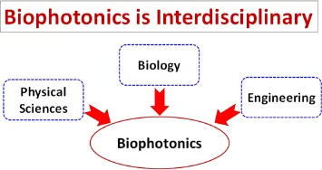
Biophotonics that combines physics, chemistry, engineering, and biology is highly interdisciplinary. At the very minimum, it is a field that brings in the physical knowledge of optical devices, such as light sources, detectors, and cameras, as well as special instruments such as the microscope to study biology. It also encompasses the knowledge of molecular structures based on our understanding of the physics and chemistry of molecules, couched in quantum mechanics. Finally, it forces us to be ever so innovative in engineering new devices and to process the massive array of data that comes with digital microscopy taken at the speed of live processes of biological significance. The illustration in Figure 1.1 points to the essential needs of all of these background expertise, and the need to truly interface these fields of study so that a new “whole” is uniquely useful for enhancing our knowledge of biology and medicine.
Indeed, the timely award of the 2014 Nobel Prize in Chemistry to three pioneering researchers, William Moerner, Eric Betzig, and Stefan Hell, in the focus area of biophotonics stands tall in helping to claim that this field has finally arrived. In each of these three cases, the idea is to be able to visualize the biological system under investigation at a hitherto unheard-of level of spatial precision and to do this while the molecules are in as native a state of function as possible. These techniques of super-resolution optical microscopy are now leading the way to our understanding of the details of molecular function within a viable cell.
Simultaneously, the Nobel Prize for Physics in 2014 was awarded to Isamu Akasaki, Hiroshi Amano, and Shuji Nakamura for their invention of the efficient blue LEDs. This discovery has led the way to new engineering approaches in designing light sources to excite fluorophores efficiently. Albert Einstein developed the theory of light emission and absorption based on the now famous “A-B” rate coefficients in 1915. Though this discovery did not net him a Nobel Prize, we celebrated 2015 as the International Year of Light worldwide. The American Physical Society and the Biophysical Society had taken advantage of the year 2015 to proclaim the strong association of optical investigation with the significant, outstanding biophysical problems. The Congress on Laser and Electro-Optics (CLEO) had arranged a special symposium where not only these distinguished scientists participated, they were joined by Steve Chu, the 1997 Nobel Laureate in Physics for his work on optical molasses and the optical atomic trap, who is actively in the field of biophotonics.
Figure 1.1. An overview of interdisciplinary science of biophotonics.
In guiding this transition and spearheading the efforts to make unique contributions in the new field of biophotonics, the United States National Science Foundation (NSF) had funded the establishment of a Center for Biophotonics Science and Technology at the University of California, Davis, creating an interface between the Colleges of Engineering, Mathematics, Physical Sciences and Biological Sciences along with the Schools of Medicine and Veterinary Medicine. This center flourished from 2002–2012 and has now evolved into a new Center for Biophotonics at the University of California, Davis. Further driving this effort is the creation of the Biophotonics World website (https://www.biophotonics.world/), linking the scientific efforts of biophotonics research worldwide.
1.2What are Photonic Processes?
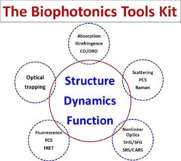
Photonic processes are those interactions whereby electromagnetic disturbance, in the form of a wave or particulate, renders changes in the material’s state and/or in the state of the incident electromagnetic radiation. We call these processes light-matter interactions. Because it is indeed the change in either the state of the matter or light that will inform us of the existence of such an interaction, we have strived to improve our approach to increase sensitivity for such detection, and in that process, gain new information about the matter being probed. So we consider the photonic methods as our “Biophotonics Tools Kit” (Figure 1.2).
Our toolkit includes, first of all, approaches to detect structures of matter (or molecules) by using just a single photon, capitalizing on the fact that electromagnetic radiation is a planar, transverse wave with specified polarization directions. These one-photon processes include absorption and birefringence; both linear and circularly polarized sources can provide unique information on the structure of matter being probed.
Figure 1.2. Biophotonics toolkit to understand structure and dynamics in biological processes.
Perhaps the most widely used light-matter interactions are those where two photons are involved. These may be of the phase-coherent processes such as light scattering, or phase-randomized, two-photon processes called fluorescence or phosphorescence. Unique identification of molecules can be achieved by designing fluorophores that have specific affinity for a specific molecule of interest. Indeed a major advance in fluorophore design was achieved by Roger Tsien, Osamu Shimomura, and Martin Chalfie who found that the green fluorescent protein (GFP) of the jellyfish (Aequorea Victoria) can be transfected into other organisms, thus rendering specific molecules of any organism fluorescently green. For this effort, they won the Nobel Prize in Chemistry in 2008.
Light scattering has been a standard-bearer for light-matter interaction studies since the time of Lord Rayleigh. Particle size differentials can easily be measured by light scattering methods, providing us with laymen arguments of why the sky is blue and a glass of milk is white when neither absorbs light. Peter Debye, who won the Nobel Prize in Chemistry in 1936 for his work on the scattering of short-wavelength light (X-rays) by molecules as a probe of molecular structure, continued to use the scattering of visible light for probing larger polymer structures. Going further into molecular spectroscopy, C.V. Raman discovered that molecular motion in the form of normal mechanical modes could create modulation signatures on the incident light shined onto the probed material, thus making these signatures the hallmark for the presence of specific molecules. For this discovery, he was awarded the Nobel Prize in Physics in 1930.
As is well-known, Charles Townes shared the 1964 Nobel Prize in Physics with Nicolay G. Basov and Aleksandr M. Prokhorov for fundamental work in quantum electronics, the fundamental precursor to the discovery of the laser. Equally important is the awarding of the Nobel Prize in Physics in 1981 to Arthur Schawlow for laser spectroscopy, shared by Nicolaas Bloembergen and Kai Siegbahn. In particular, Bloembergen started a new field of physics called nonlinear optics, whereby intense laser fields can elicit non-linear materials responses, producing signals that are unique to the nature of the interaction and that of the material. This opened up the ability to capitalize these relatively weak material processes to identify material specificity more uniquely. We shall delve into nonlinear optics and related nonlinear optical microscopy in this volume.
Rounding out this toolbox, it is indeed the pioneering work of Steve Chu and colleague, Arthur Ashkin, that evolved the atomic trap into a tool for trapping larger molecules and for measuring forces at the molecular scale (pico-Newtons). This process depends on the radiation pressure gradient that can be exerted onto a well-controlled shaped particle of nearly micron size. Using this technique biologists are beginning to develop quantitative biological schemes of measurement, connecting force at this molecular scale to functions and structures.
With all these biophotonics tools in the toolbox of light, it is now time to move on to the next step and examine biological processes that can benefit from these tools.
1.3Fundamental Biology Studies Need Photonics
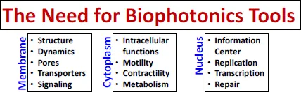
In its most rudimentary view, biology describes an organism that has spatial extent, consumes matter for its chemical content, utilizes this chemical content by converting it into energy for the purpose of cellular metabolism, excretes waste matter, and is dynamic. We now know much more about the details of an organism, even one as simple as a single-celled bacterium or algae, or even a virus. Principally, the fundamental unit of the living entity is the cell. The cell, however, is not just a blob but is composed of millions of molecules each serving some special function to maintain life for the cell. As the technology in light microscopy improved, more and more details of a cell have become evident. Being true scientists, inquisitive individuals of the earlier era have been able to identify regions of the live cell and to assign differential functionality to them.
Cells all have a protective surface. Optically, using a transmission microscope of relatively low spatial resolution (e.g., 40×), one can show that the region of the cell cover is definitely distinct from that of the cell interior. Furthermore, in all eukaryotic cells, there are dense bodies within the interior of the cell. These are called nuclei. These cells are often dynamic in shape, frequently contorting themselves into highly convoluted shapes. The nuclear content of cells have become associated with genetic material, and the composition of the nucleus has been identified as deoxyribonucleic acid or DNA. By using another photonic method, X-ray diffraction, James D. Watson, Francis Crick and Maurice Wilkins (winners of the Nobel Prize in Physiology or Medicine in 1962) deciphered that the DNA within the cell consists of a double helix structure. Characterizing the DNA as a set of fundamental building blocks, composed of only four different DNA molecules, Adenine (A), Thymine (T), Guanine (G), and Cytosine (C), allowed scientists to postulate the origin of genetic transcription. The finding that in a double helix, the pairing of A–T and G–C is inviolable led to our understanding of how the double helix functions in the complementary template pairing scheme for the preservation of genetic material during transcription and cell division processes. Other optical methods, the optical rotatory dispersion and circular dichroism, allowed scientists to speculate how molecular melting can occur, leading to genetic mutations during cell division. Just how these activities take place in the cell and in the nucleus requires a higher spatial resolution optical microscope. Indeed, photons extending more into the X-ray wavelength domain play an important role in ascertaining that not only do proteins exist within the cell, but they also have major functions both on the cellular surface (membrane) as well as in the cell nucleus. Today we know many of these to be proteins. Linus Pauling’s work on deciphering the molecular structures of proteins using X-ray diffraction methods won him the 1954 Nobel Prize in Chemistry. Now we know that there are many types of special proteins: membrane proteins, nuclear proteins, as well as proteins that self-assemble within the cytoplasm to form definitive molecular structures, foretelling the anticipated shapes and functions of the cells.
Under optical microscopic observation, with a resolution of approximately 1.0 micrometer (µm), cells of different shapes can be ascertained. The time trace of live cells presents another problem: Why do the cells change shape? They have been known to change shape in search of nutrients from external sources. The process of cytokinesisdescribes the movement of a cell. Voluntary and highly ordered rearrangement of proteins within a cell occurs in muscle cells upon a trigger signal, electrical or chemical. This suggests a sensory network within the cell for cell signaling and transduction. Molecular sources of such organized cellular activity in muscle cells have been identified as Ca++ ions. By using another optical method, fluorescence emission, it became possible to identify the movement of the Ca++ flux during nerve cell signaling processes. Although higher spatial resolution can be achieved by the use of electron microscopy (EM), the preparation protocol in EM usually precludes studying the cell domain in their functional state. Recent developments in cryo-EM have enhanced the value of this technique immensely. Appropriately, researchers of this technique development have been awarded the 2017 Nobel Award in Chemistry. Given the charge to the current surge of investigators that it would be most desirable to examine biological systems in their native functional state, it is the probing of the dynamics of the intracellular structure as they relate to functions of the cell that the field of biophotonics is tackling.
Where are the photonic needs? First of all, the cell membrane is composed of lipid molecules arranged into a molecular bilayer. This structure has a typical depth of approximately 5 nanometers (nm). The protein structures that dot the landscape of the membrane often are of dimensions of approximately 10 nm, and the membrane structure is now known to be highly heterogeneous. There is also the fact that the heterogeneity in the structure is highly dynamic, consistent with the dynamic movement (diffusion in two dimensions) on the membrane. The protein composition within the cytoplasm is completely dynamic; hardly is there a moment that a static structure can be ascertained. Even in the driven movements of collective muscle proteins, the head groups of the molecule within the myosin subfragment-I (S-1) that is the molecular motor is always in motion unless the cell is in the rigor state. Within the cell nucleus, nuclear proteins are constantly in search of DNA defects, in an effort to “search and destroy” before any defective genetic domain manifests as mutated genetic materials. How do we probe any and all of these functionalities of the cell? These are the outstanding types of questions that exist spanning the entire spectrum of biological molecules within a cell. As optical methods have evolved rapidly over the last 50 years, the application of new photonic methods to biology, hence biophotonics, becomes crucial for furthering the understanding of the dynamic assemblies of molecular networks within a cellular environment (Figure 1.3).
Figure 1.3. The role of biophotonics tools to understand the molecular cell.
1.4Applied Biology — Molecular Medicine Using Photonic Means
As humankind continues to inhabit the earth, the health of each continues to play a major role in the perpetuation of individuals and species. Healthcare in any society constitutes one of the largest components of that society’s budget, and one is finding out that diseases ar...