Essentials of Ophthalmology
eBook - ePub

Essentials of Ophthalmology

For Medical School and Beyond

  1. 272 pages
  2. English
  3. ePUB (mobile friendly)
  4. Available on iOS & Android
eBook - ePub

Essentials of Ophthalmology

For Medical School and Beyond

About this book

Essentials of Ophthalmology: For Medical School and Beyond is a concise reference text for the medical undergraduates and residents, but the contents will also benefit family physicians who really are the first line of eye-care givers in the community.

Highly illustrated with 200 colour clinical pictures and illustrations, the book is written by the multi-disciplinary faculty of the Department of Ophthalmology, National University Health System, Singapore. The authors constantly asked themselves 'how much is too much' and deliberately attempted to weed out any 'excess' for the benefit of the target readers. The content is written in a point format with lucid language.

Emphasis has been focused on information that constitutes essential ophthalmic 'core conditions and problems' of the current medical undergraduate curriculum. Every section in the book has 'learning objectives' and a 'take home message' to facilitate quick learning. The book embraces a practical guide to the study of ocular diseases, basic methods of investigations and treatment where applicable.


Contents:

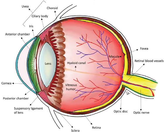
  • Basic Anatomy of the Eye, Adnexa and Visual Pathways (Jeyabal Preethi, Koh Teck Chang Victor)
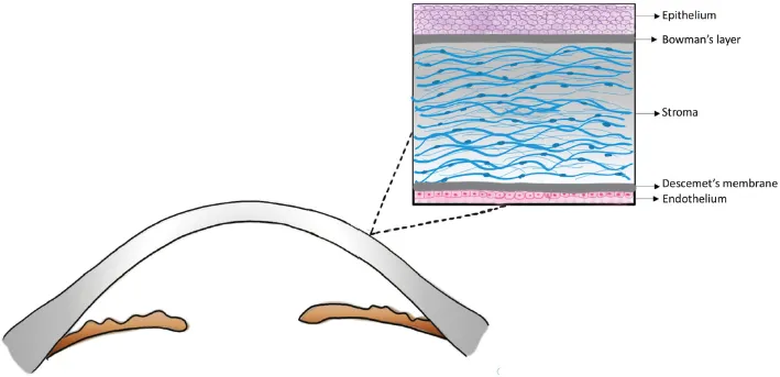
  • Cornea and External Eye Diseases (Chai Hui Chen Charmaine, Anna Marie Tan Wee Tien, Ray Manotosh)
  • Cataract (Chai Hui Chen Charmaine, Anna Marie Tan Wee Tien, Ray Manotosh)
  • Glaucoma (Koh Teck Chang Victor, Chen Ziyou David)
  • Uveitis (Chan Hwei Wuen, Dawn Lim Ka-Ann)
  • Vitreoretinal Disorders (Yuen Yew Sen, Paul Zhao Song Bo)
  • Oculoplastics (Stephanie Ming Young, George Thomas Naveen, Shantha Amrith, Gangadhara Sundar)
  • Paediatric Ophthalmology and Strabismus (Lam Sing Harn Janice, Cheryl Ngo Shufen)
  • Neuro-Ophthalmology (Lin Hui'en Hazel Anne, Clement Tan Woon Teck)
  • Principles and Practice of Low Vision Rehabilitation (Daniel Bohan)
  • Clinical Approaches:
    • Approach to Leukocoria (Lam Sing Harn Janice)
    • Clinical Pathway for Chronic Visual Loss (Jeyabal Preethi, Koh Teck Chang Victor)
    • Clinical Pathway for Acute Visual Loss (Lin Hui'en Hazel Anne)
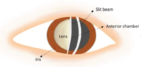
    • Diagnostic Flowchart in an Acute Red Eye (Chai Hui Chen Charmaine, Anna Marie Tan Wee Tien, Ray Manotosh)
    • Neuro-ophthalmology Approaches (Lin Hui'en Hazel Anne)
    • Pupil Examination (Lin Hui'en Hazel Anne)
  • Clinical Examinations:
  • Approaches to Visual Acuity (Jeyabal Preethi, George Naveen Thomas)
  • Cover Test (Lam Sing Harn Janice)
  • Normal Fundus Examination (Yuen Yew Sen)
  • Visual Fields by Confrontation (Lin Hui'en Hazel Anne)


Readership: Medical undergraduates, residents, primary eye-care providers.
Key Features:

  • The book is designed to incorporate knowledge and skills to prepare the undergraduate medical students for their NUS rotation within Ophthalmology
  • The content is written in summarized point format with lucid language that is supplemented with more than 200 high resolution clinical pictures and illustrations
  • Atlas of coloured images which illustrates concepts, anatomy, conditions, treatment and common complications related to the Asian population

Frequently asked questions

Yes, you can cancel anytime from the Subscription tab in your account settings on the Perlego website. Your subscription will stay active until the end of your current billing period. Learn how to cancel your subscription.
No, books cannot be downloaded as external files, such as PDFs, for use outside of Perlego. However, you can download books within the Perlego app for offline reading on mobile or tablet. Learn more here.
Perlego offers two plans: Essential and Complete
  • Essential is ideal for learners and professionals who enjoy exploring a wide range of subjects. Access the Essential Library with 800,000+ trusted titles and best-sellers across business, personal growth, and the humanities. Includes unlimited reading time and Standard Read Aloud voice.
  • Complete: Perfect for advanced learners and researchers needing full, unrestricted access. Unlock 1.4M+ books across hundreds of subjects, including academic and specialized titles. The Complete Plan also includes advanced features like Premium Read Aloud and Research Assistant.
Both plans are available with monthly, semester, or annual billing cycles.
We are an online textbook subscription service, where you can get access to an entire online library for less than the price of a single book per month. With over 1 million books across 1000+ topics, we’ve got you covered! Learn more here.
Look out for the read-aloud symbol on your next book to see if you can listen to it. The read-aloud tool reads text aloud for you, highlighting the text as it is being read. You can pause it, speed it up and slow it down. Learn more here.
Yes! You can use the Perlego app on both iOS or Android devices to read anytime, anywhere — even offline. Perfect for commutes or when you’re on the go.
Please note we cannot support devices running on iOS 13 and Android 7 or earlier. Learn more about using the app.
Yes, you can access Essentials of Ophthalmology by Ray Manotosh, Victor Koh in PDF and/or ePUB format, as well as other popular books in Medicine & Anatomy. We have over one million books available in our catalogue for you to explore.

Information

Publisher
WSPC
Year
2018
eBook ISBN
9789813275614
Subtopic
Anatomy

Chapter 1

BASIC ANATOMY OF THE EYE, ADNEXA AND VISUAL PATHWAYS

1.1Basic Anatomy

Learning Objectives
Understand the basic anatomical structures of the globe.
The external structure of the globe comprises the sclera (outer most layer), uveal tissue (middle layer) and retina (innermost layer). Refer to Fig. 1.1.
Sclera
Strong and dense coat protecting the intraocular contents
Anterior 1/6th — transparent cornea, posterior 5/6th opaque sclera. Junction is called limbus
figure
Fig. 1.1. Coronal section of the eyeball showing the various structures.
Thickness:
figure
Thickest posteriorly (1 mm) and gradually becomes thinner upon tracing anteriorly
figure
Thinnest at the level of insertion of extraocular muscles
Lamina cribrosa: sieve-like part of sclera through which optic nerves exits the globe
Uveal tissue
Supplies nutrition to various structures of the eyeball
From anterior to posterior, it consists of iris, ciliary body and choroid
Functions:
figure
Iris: iris colour varies among different individuals depending on the amount of melanin. Controls amount of light entering the eye
figure
Ciliary body: aqueous humour production, accommodation
figure
Choroid: supplies oxygen and nutrition to outer layers of retina
Retina
See the sections below for more details
Take home messages
The cornea and sclera forms the outermost wall of the globe and forms a protective cover over the intraocular structures.

1.2Cornea

Learning Objectives
Understand the components and functions of the different layers of the cornea.
The cornea is the transparent outermost covering of the eye and is the most important refractive medium of the eye (Fig. 1.2 and Fig. 1.3).
figure
Fig. 1.2. Illustration of slit-lamp beam image of the anterior segment. The first broad slit beam corresponds to the cornea and the second narrow beam corresponds to the anterior surface of the lens.
figure
Fig. 1.3. Cross-section of the cornea and the 5 layers.
The 5 layers of the cornea are composed of:
Epithelium
5 layers of cells and 50-60 microns thick
Approximately 7 days for complete turnover of corneal surface epithelium
Production of new cells occurs at the limbus and grows centripetally from the periphery towards the centre
Nerve endings of sensory nerve fibres run between the epithelial cells
Bowman’s Layer
Acellular layer
8–12 microns thick
Stroma
Forms 90% of corneal thickness
Multiple lamellae of compact collagen fibrils
Uniform spacing of collagen fibrils maintain the transparency
Descemet’s Membrane
Basement membrane of the endothelium
10 microns thick
Endothelium
Single layer of flattened cells
Important in the transport of fluid, keeping the cornea dehydrated and transparent
Does not regenerate
Physiological rate of loss of cells with age

Nerve Supply of the Cornea

From the ophthalmic division of the trigeminal nerve (mainly...

Table of contents

  1. Cover Page
  2. Title Page
  3. Copyright Page
  4. Contents
  5. Foreword
  6. Preface
  7. Acknowledgements
  8. About the Editors
  9. List of Contributors
  10. Chapter 1 Basic Anatomy of the Eye, Adnexa and Visual Pathways
  11. Chapter 2 Cornea and External Eye Diseases
  12. Chapter 3 Cataract
  13. Chapter 4 Glaucoma
  14. Chapter 5 Uveitis
  15. Chapter 6 Vitreoretinal Disorders
  16. Chapter 7 Oculoplastics
  17. Chapter 8 Paediatric Ophthalmology and Strabismus
  18. Chapter 9 Neuro-Ophthalmology
  19. Chapter 10 Principles and Practice of Low Vision Rehabilitation
  20. Chapter 11 Clinical Approaches
  21. Chapter 12 Clinical Examinations
  22. Glossary
  23. Index