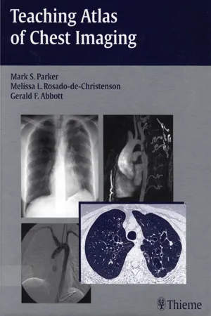
Teaching Atlas of Chest Imaging
- 800 pages
- English
- PDF
- Available on iOS & Android
Teaching Atlas of Chest Imaging
About this book
This lavishly illustrated book is your comprehensive, hands-on guide to evaluating chest images. It is ideal for reading cover-to-cover, or as a reference of radiological presentations for common thoracic disorders. With this book, you will learn to interpret chest images and recognize the imaging findings, generate an appropriate differential diagnosis, and understand the underlying disease process.
The atlas begins with a review of normal thoracic radiography, CT, and MR anatomy, and goes on to present cases on a wide range of congenital, traumatic, and acquired thoracic conditions. Each case is supported by a discussion of etiology, pathology, imaging findings, treatment, and prognosis in a concise, bullet format to give you a complete clinical overview of each disorder. More than 1, 050 high-quality images demonstrate normal and pathologic findings, and complementary scans demonstrate additional imaging manifestations of disease entities.
Residents, fellows, and general radiologists called upon to interpret chest images will find this easy-to-use book invaluable as a learning tool and reference. It is also a must for thoracic radiologists, pulmonary physicians, and thoracic surgeons who must read chest images—especially of challenging cases.
Frequently asked questions
- Essential is ideal for learners and professionals who enjoy exploring a wide range of subjects. Access the Essential Library with 800,000+ trusted titles and best-sellers across business, personal growth, and the humanities. Includes unlimited reading time and Standard Read Aloud voice.
- Complete: Perfect for advanced learners and researchers needing full, unrestricted access. Unlock 1.4M+ books across hundreds of subjects, including academic and specialized titles. The Complete Plan also includes advanced features like Premium Read Aloud and Research Assistant.
Please note we cannot support devices running on iOS 13 and Android 7 or earlier. Learn more about using the app.
Information
Table of contents
- Teaching Atlas of Chest Imaging
- Title Page
- Copyright
- Dedication
- Contents
- Foreword
- Preface
- Acknowledgments
- Contributors
- SECTION I: Normal Thoracic Anatomy
- SECTION Il: Developmental Anomalies
- SECTION III: Airways Disease
- SECTION IV: Atelectasis
- SECTION V: Pulmonary Infections and Aspiration Pneumonia
- SECTION Vl: Neoplastic Diseases
- SECTION VII:Trauma
- SECTION VIII: Life Support Tubes, Lines, and Monitoring Devices
- SECTION IX: Diffuse Lung Disease
- SECTION X: Occupational Lung Disease
- SECTION XI: Adult Cardiovascular Disease
- SECTION XII: Abnormalities and Diseases of the Mediastinum
- SECTION XIII: Pleura, Chest WaII, and Diaphragm
- INDEX