eBook - ePub
Natural Therapies for Emphysema and COPD
Relief and Healing for Chronic Pulmonary Disorders
- 208 pages
- English
- ePUB (mobile friendly)
- Available on iOS & Android
eBook - ePub
Natural Therapies for Emphysema and COPD
Relief and Healing for Chronic Pulmonary Disorders
About this book
The first book to address emphysema and chronic obstructive pulmonary disease (COPD) from a nutritional and alternative medicine approach
• Explains the benefits of detoxification, dietary changes, and food combining
• Details 45 suggested herbs and 26 nutritional supplements as well as information on how to stop smoking
Approximately 35 million people in the United States have been diagnosed with some form of chronic obstructive pulmonary disease (COPD)--emphysema constituting 18 million of that group. Worldwide, as many as 293 million people suffer with these conditions. COPD is the fourth leading cause of death in America, claiming nearly 120,000 lives annually. Yet conventional approaches to treatment, with their regimens of drugs and unceasing physical therapy, provide neither cure nor significant relief.
In Natural Therapies for Emphysema and COPD, Robert Green shows that alternative holistic therapies ranging from herbs to homeopathy offer great promise in relieving COPD’s debilitating symptoms. Starting with the basics of the physiology of respiration, Green presents a comprehensive program that includes detoxification, dietary changes, nutritional supplements, and herbal medicine; breathing techniques and exercise options such as aerobics, yoga, qigong, and tai chi; and alternative therapies such as homeopathy, acupuncture, and massage--noting how and why each therapy works. He also details how to stop smoking, includes resources for alternative health practitioners, and provides sources for the alternative products recommended.
Frequently asked questions
Yes, you can cancel anytime from the Subscription tab in your account settings on the Perlego website. Your subscription will stay active until the end of your current billing period. Learn how to cancel your subscription.
No, books cannot be downloaded as external files, such as PDFs, for use outside of Perlego. However, you can download books within the Perlego app for offline reading on mobile or tablet. Learn more here.
Perlego offers two plans: Essential and Complete
- Essential is ideal for learners and professionals who enjoy exploring a wide range of subjects. Access the Essential Library with 800,000+ trusted titles and best-sellers across business, personal growth, and the humanities. Includes unlimited reading time and Standard Read Aloud voice.
- Complete: Perfect for advanced learners and researchers needing full, unrestricted access. Unlock 1.4M+ books across hundreds of subjects, including academic and specialized titles. The Complete Plan also includes advanced features like Premium Read Aloud and Research Assistant.
We are an online textbook subscription service, where you can get access to an entire online library for less than the price of a single book per month. With over 1 million books across 1000+ topics, we’ve got you covered! Learn more here.
Look out for the read-aloud symbol on your next book to see if you can listen to it. The read-aloud tool reads text aloud for you, highlighting the text as it is being read. You can pause it, speed it up and slow it down. Learn more here.
Yes! You can use the Perlego app on both iOS or Android devices to read anytime, anywhere — even offline. Perfect for commutes or when you’re on the go.
Please note we cannot support devices running on iOS 13 and Android 7 or earlier. Learn more about using the app.
Please note we cannot support devices running on iOS 13 and Android 7 or earlier. Learn more about using the app.
Yes, you can access Natural Therapies for Emphysema and COPD by Robert J. Green in PDF and/or ePUB format, as well as other popular books in Medicine & Alternative & Complementary Medicine. We have over one million books available in our catalogue for you to explore.
Information


Essential Respiratory Anatomy and Physiology
The respiratory system is a marvelously complex system that maintains one of the most vital functions of the human body: breathing. The well- being of the entire body is vitally dependent upon proper functioning of every detail of respiration—the process by which the body takes in oxygen, distributes oxygen to cells throughout the body, and releases carbon dioxide created as a by-product of the process. The respiratory system must not only operate efficiently, it must also protect itself from environmental irritants and infection. The main purpose of the lungs is to facilitate the exchange of oxygen from the air we breathe with carbon dioxide given off by the cells of the body. The blood circulation transports oxygen to the cells of the body and carries carbon dioxide back to the lungs to be exhaled in the breath. Thus, the heart and circulatory system also play a vital role in respiration.
Respiration is a four-phase process. The first phase of respiration is breathing, otherwise known as pulmonary ventilation. Breathing consists of inspiration (inhaling) and expiration (exhaling) of air. This vital function is controlled by brain structures called the medulla oblongata and the pons. These structures, which are located in the brain stem portion of the central nervous system, send nerve impulses that stimulate the muscles that control the breathing process (the thoracic muscles and the diaphragm). The nervous system control of breathing is predominantly an involuntary (automatic) process, but can be voluntarily controlled as needed. The voluntary aspect is best evidenced in the interruption of breathing when you cough or when you hold your breath, for example.
In the second phase of respiration, technically known as pulmonary or external respiration, an exchange of oxygen and carbon dioxide gases occurs between the lungs and the blood. Oxygen is transferred from the lungs into the blood to be distributed to tissues all over the body. At the same time, carbon dioxide is removed from the blood to be exhaled in the breath. Efficient oxygen transfer depends on the presence of sufficient amounts of hemoglobin in the red blood cells, which are responsible for carrying oxygen from the lungs to the tissues. This is why individuals who are anemic, having lower than optimal amounts of hemoglobin, have additional concerns that complicate their COPD issues.
The third phase of respiration, which does not take place in the lungs, is called internal (tissue) respiration. This phase involves the exchange of gases between the blood and the tissues of the body.
The fourth and final phase of respiration, cellular respiration, occurs inside the body’s cells. Through the process of cellular respiration, the body uses the oxygen that has been obtained from the blood to make energy and sustain metabolic activity. As part of this process, the body generates carbon dioxide as a waste product.
GENERAL ANATOMY OF THE LUNGS
The lungs are elastic, spongelike organs that lie in the thoracic cavity (the chest). The lungs are enclosed within the rib cage. The top of the lungs reaches slightly above the clavicle (collarbone), and the base of the lungs, which is slightly curved, fits over the diaphragm (a muscular structure that separates the chest cavity from the abdominal cavity). The right lung is somewhat shorter than the left to accommodate the space needed by the liver, which lies just below the diaphragm under the right lung.
A two-layered membrane composed mainly of elastic tissue (the pleural membrane) encloses and protects the lungs within the thoracic cavity. The inner membrane, called the visceral pleura, lines the lungs themselves. The outer membrane, the parietal pleura, is attached to the wall of the thoracic cavity. A thin film of fluid (called pleural fluid) occupies the space between the two membranes in order that their surfaces can easily slide over one another during inspiration and expiration.
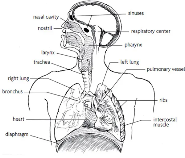
Fig. 1. The respiratory system and related structures.
The right lung consists of three lobes and the left lung has two. The right lung is also divided into ten sections and the left into nine sections. These sectional subdivisions amount to what can be thought of as a three-dimensional map that allows for greater precision in specifying locations within the lungs.
PATHWAY OF AIRFLOW THROUGH THE RESPIRATORY SYSTEM
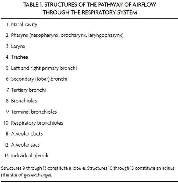
The respiratory system begins at the nostrils, where air enters the nasal cavity and proceeds into the nasopharynx (the passage that connects the nose with the throat). As air passes through the structures of the nasal cavity, it is filtered by coarse nasal hairs and warmed and humidified by a highly vascularized mucous membrane. From the nasopharynx, the air continues down the oropharynx and the laryngopharynx (the back of the mouth and the throat) through the larynx (voice box) into the trachea (windpipe). The trachea is a tube constructed of cartilage that extends down from the larynx about four inches. At this point, it divides into two branches, called the primary bronchial tubes. These—the right and left primary bronchi—are the first two main branches of the bronchial tree.
Where the primary bronchi enter the lungs, they begin branching into smaller bronchi called secondary or lobar bronchi. These secondary bronchi divide and branch into tertiary bronchi, which further divide into even smaller tubes called bronchioles, which themselves continue to divide into smaller branches. Bronchioles eventually become tiny terminal bronchioles that give rise to branches of respiratory bronchioles. These respiratory bronchioles ultimately end in clusters of air sacs that contain individual alveoli.
Alveoli are tiny air-containing cavities at the endpoint of the respiratory passageways in the lungs. Each lung has roughly 300 million alveoli and each one of them is essentially a gas bubble that is surrounded by a network of capillaries (tiny blood vessels).
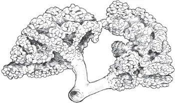
Fig. 2. Two acini, showing a terminal bronchiole splitting into two respiratory bronchioles with alveolar ducts and alveoli.
Alveoli are connected to the respiratory bronchioles by passageways called alveolar ducts. Two or more alveoli that share a common alveolar duct are called alveolar sacs.
Terminal bronchioles along with their respective respiratory bronchioles and alveolar sacs are known as lobules. Respiratory bronchioles and their alveolar sacs are collectively known as the acinus. The acinus is essentially a lobule without the terminal bronchiole, and it is where actual gas exchange in the lungs occurs. The acinus is something like a cluster of grapes, in which the main stem coming off the vine is the respiratory bronchiole, the smaller stems are the alveolar ducts, and the grapes themselves represent the alveoli.
The alveoli are separated from one another by membranes called the interalveolar septa (a matrix consisting of connective tissue fibers and capillaries), and are connected to each other by what are known as pores of Kohn. These pores are essentially holes that exist between the alveoli that allow for the complete ventilation of air throughout the acinus. It is essential to realize that there is no truly free space between the alveoli. They are always surrounded by a matrix of connective tissue fibers and capillaries—the interalveolar septum.
STRUCTURAL SUPPORT OF THE RESPIRATORY SYSTEM
From the trachea to the bronchioles, the respiratory tract is supported primarily by rings and plates made of cartilage (tough, elastic tissue). The cartilage disappears in the bronchioles, as bronchioles themselves are not supported by cartilage, but rather are surrounded by smooth muscle, which allows for fluctuation in the size of the bronchiole. Bronchioles are also characterized by the presence of elastic fibers that surround them in addition to the smooth muscle.
BLOOD SUPPLY TO THE LUNGS
There are two separate systems of blood flow in the lungs: pulmonary circulation and bronchial. The first of these two systems, pulmonary circulation, involves the blood that circulates from the heart through the pulmonary blood vessels. This is the part of the circulatory system that picks up oxygen from the lungs, transports it to the tissues throughout the body, and returns the blood back to the lungs after the oxygen has been depleted by the tissues. The returning blood contains carbon dioxide excreted by the tissues as a waste product of metabolism.
As oxygen-rich blood circulates from the lungs throughout the body, the tissues take the oxygen they need from the blood. Thus the blood that is returned to the heart is essentially without oxygen. The heart sends this deoxygenated blood to the lungs by way of the pulmonary artery, through which it reaches the capillaries that surround the alveoli. Here, at the interface between the alveoli and capillaries, the blood receives a fresh supply of oxygen. After passing through the alveolar capillaries, the freshly oxygenated blood makes its way back to the heart by way of the pulmonary vein, where it then exits the heart through the aorta (a major artery) and circulates through another cycle of delivering oxygen to the tissues of the body. Upon receiving this fresh supply of oxygen, the tissues are now able to take care of their metabolic business. Carbon dioxide is one of the waste products of metabolism and the tissues will deliver carbon dioxide back into the bloodstream, where it will be carried back to the lungs and exhaled out of the body.
You may have learned that arteries are blood vessels that carry oxygen-rich blood away from the heart to the rest of the body and veins are blood vessels that carry oxygen-poor blood from the tissues back to the heart. This is mostly correct. However, the definition of an artery or vein actually hinges on the direction of blood flow toward or away from the heart, not the oxygen content of the blood the vessel carries. The pulmonary artery, for example, carries deoxygenated blood away from the heart. The pulmonary vein, which transports oxygen-rich blood from the alveolar capillaries in the lungs, is still a vein because it is taking blood toward the heart.
The second system of blood flow in the lungs, bronchial circulation, involves the circulation of blood through the blood vessels that nourish the lung tissue itself. The bronchial arteries receive oxygenated blood mainly from the aorta, and deliver this oxygen-rich blood to the lung tissue (e.g., the bronchi and bronchioles and the bronchial lymph nodes) via the capillaries adjacent to the cells of these tissues. After the lung tissue takes the oxygen it needs from the bronchial arteries, some of the now deoxygenated blood is returned to the heart by way of the bronchial veins and some by way of the pulmonary veins.
LINING OF THE RESPIRATORY TRACT AND THE ALVEOLI
The respiratory tract is lined by a membrane called the respiratory epithelium. Epithelium is one of the four primary tissue types of the body. Epithelium lines the skin as well as internal organs and hollow cavities such as the intestines and the respiratory passageways. Epithelial cells line the insides of the lungs. The nasal cavity, nasopharynx, and other parts of the respiratory system (from the larynx to the bronchioles, but not including the vocal cords) are lined by a membrane containing epithelial cells with many mucus-secreting goblet cells. There are also many other mucus-secreting glands throughout the walls of the trachea and the bronchi, but not the bronchioles. These cells and glands help “condition” inhaled air to keep the respiratory system clean and free of particles.
Moist mucus secreted by these cells and glands traps foreign matter. At the same time, cilia (tiny hairlike structures) on the epithelium sweep away particles so that they may be swallowed or expectorated. This is the natural course of action for maintaining the respiratory passageways. Water to humidify inhaled air is derived from mucus, and heat to warm the air comes from the abundant underlying blood vessels. These processes work together to ensure that the air that travels through the respiratory tract is not only at body temperature, but also as clean as possible, and completely humidified.
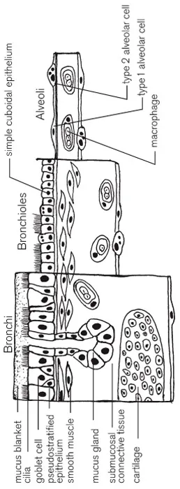
Fig. 3. The overall changes in the structure of the lining of the respiratory tract.
From the bronchioles onward, the branching network of the lungs becomes very extensive, and the nature of the cells lining the respiratory tract gradually begins to change. Once we reach the terminal bronchioles, the cells lining the respiratory tract no longer produce mucus, nor do they have cilia, and an immune system cell known as a macrophage assumes the responsibility of removing inhaled foreign particles. Rather than trapping and sweeping away foreign matter like cilia, macrophages engulf and digest the particles. As the terminal bronchioles eventually become respiratory bronchioles, the cells lining the respiratory tract change yet again into a cell type known as squamous epithelium, the same kind of cells that line the alveolar wall.
The walls of the alveoli are composed of only a single layer of cells, and they lie immediately next to capillaries that also consist of only a single layer of cells. The alveolar wall also contains its own macrophages, which are responsible for removing dust particles and other debris from the alveoli. Because the inner surface of the alveoli is moist and comes in direct contact with air (gas), the watery surface of the inner alveolar wall is always attempting to contract—much in the way water holds itself together on the rim of a glass that is about to overflow. This contracting force exhibited by the watery inner alveolar surface makes for a naturally existing surface tension at the liquid–gas interface of the inner alveolar wall. In effect, this tension inadvertently forces air out of the alveoli through the bronchioles, thus promoting the collapse of the alveoli. To compensate for this phenomenon, certain alveolar cells secrete a fluid that contains a surfactant that lowers the surface tension of the liquid– gas barrier and prevents the alveoli from collapsing upon exhalation.
GAS EXCHANGE IN THE ALVEOLI
Successful exchange and efficient transport of oxygen and carbon dioxide are critical to sustaining the immediate cellular requirements of life. In one way or another, emphysema, chronic bronchitis, bronchiectasis, and the complications of infections and pneumonia all obstruct the airway and compromise the effectiveness of gas exchange. Many of the problems associated with emphysema occur in the ...
Table of contents
- Cover Image
- Title Page
- Dedication
- Epigraph
- Acknowledgments
- Table of Contents
- Introduction
- Chapter 1: Essential Respiratory Anatomy...
- Chapter 2: Understanding COPD and Emphysema
- Chapter 3: Diagnosis and Conventional...
- Chapter 4: Quitting Smoking
- Chapter 5: Dietary Therapeutics
- Chapter 6: Dietary Supplements
- Chapter 7: Herbal Medicine
- Chapter 8: Exercise, Breathing Techniques,...
- Chapter 9: Other Alternatives and...
- Appendix 1: Additional Resources
- Appendix 2: Recommended Reading
- Glossary
- Selected Bibliography
- About the Author
- About Inner Traditions • Bear & Company
- Books of Related Interest
- Copyright & Permissions