Mycobacteria are bacterial pathogens which cause diseases in humans and non-human animals. This monograph primarily covers the most important and widely researched groups of mycobacteria: members of the Mycobacterium tuberculosis complex (MTC) and Mycobacterium leprae, across a wide range of host species. M. tuberculosis and M. bovis are particularly relevant with the increasing drug resistance and co-infection with HIV associated with M. tuberculosis and the possible cross-infection of badgers and cattle associated with M. bovis. This book provides a reference for researchers working in different fields, creating a work which draws together information on different pathogens, and by considering the diseases in a zoonotic context, provides a One Health approach to these important groups of diseases.
eBook - ePub
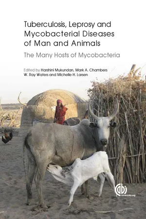
Tuberculosis, Leprosy and other Mycobacterial Diseases of Man and Animals
The Many Hosts of Mycobacteria
- English
- ePUB (mobile friendly)
- Available on iOS & Android
eBook - ePub
Tuberculosis, Leprosy and other Mycobacterial Diseases of Man and Animals
The Many Hosts of Mycobacteria
About this book
Trusted by 375,005 students
Access to over 1.5 million titles for a fair monthly price.
Study more efficiently using our study tools.
Information
Topic
MedicineSubtopic
Veterinary Medicine 1 Introduction and Epidemiology of Mycobacterium tuberculosis Complex in Humans
1National Institutes of Health, Bethesda, USA
2Yonsei University College of Medicine, Seoul, Republic of Korea

* E-mail: [email protected]
History of Tuberculosis
Tuberculosis (TB) is arguably one of the most devastating diseases that have afflicted mankind from time immemorial. Known by many different names throughout history, such as phthisis, scrofula, consumption, King’s Evil, lupus vulgaris, the white plague and ‘captain of all these men of death’, the scourge remains a significant public health concern. Perhaps the earliest evidence of TB comes from skeletal remains from burial sites from the latter part of the last Stone Age. Both macroscopic as well as microscopic evidence of TB, using modern scientific methods, has been found from excavations of mummified bodies from tombs from ancient Egypt dating as far back as 2400 BC (Allison et al., 1961; Nerlich et al., 1997; Zink et al., 2003). Drawings, pottery and statues of ancient Egypt that date up to 3000 BC have shown physical deformities that appear to show typical characteristics of TB of the spine (Vasiliadis et al., 2009; Dyer, 2010).
The first available writings about ‘phthisis’, meaning ‘wasting away’ in Greek, by Hippocrates (~460–370 BC) in his Of Epidemics dates as far back as 400 BC. Hippocrates, who is largely thought to be the father of modern medicine, believed phthisis was caused by growths in the lung, which he referred to as tubercular. He described phthisis as the most widespread disease of the era and provided detailed descriptions of the disease that included fevers, sweats, cough and wasting which closely resemble those of TB. The devastating nature of the disease even led Hippocrates to advise other physicians to avoid visiting ‘consumption’ patients with advanced disease because they would inevitably die and destroy the reputation of the attending physician. As pulmonary phthisis was commonly seen among close family members, Hippocrates and others widely considered the disease to be hereditary, a notion that persisted over a century. Aretæus, a Greek physician monk, described ‘consumption’ as ‘a disease with a poor prognosis that was characterized by a chronic discharge of opaque, whitish yellow fluid from the lungs’ (Dyer, 2010, p. 31). He associated people with a pale, slender and weak body type to be highly likely to develop TB. Another Greek physician, Clarissimus Galen (130–200 AD) downplayed the prevailing consideration of TB as a hereditary disease and instead came up with another theory that suggested transmission from person to person as another way by which TB could be spread. This alternate proposition ushered in the possibility, even at this very early stage, of an infectious nature of the disease that would ultimately be proved to be right. Later Girolamo Franscatoro (1478–1553), an Italian physician, suggested that phthisis could be transmitted by invisible particles which he called seminara, and that the disease was a result of a lung ulcer. Franscatoro was also the proponent of the use of the term phthisis to be restricted to the description of only pulmonary consumption instead of its common use that referred to all cases of ‘wasting’. The development of techniques for performing post-mortems by Andreas Vesalius (1514–1564) and his colleagues in the 16th century further advanced knowledge of TB by introducing a way in which specific symptoms could be associated with the cause of death.
The precise pathological and anatomical descriptions of the disease only began to appear in the 17th century when in 1679 a Dutch physician, Franciscus de la Boë (Sylvius), identified the ‘tubercle’ as a consistent characteristic change in the lungs and other areas of ‘consumptive’ patients. One of Sylvius’ students, Thomas Willis (1621–1675), related the localized lesions in the lungs and other organs to the general wasting away of the body. Another of his students, Richard Morton (1637–1698), described the three stages of phthisis: initial inflammation, formation of tubercles, and progression to ulcers and fully fledged consumption disease. Together, Willis and Morton described a form of TB that affected lymph nodes in the neck, which they called scrofula. In 1702, Mange went on to describe the pathological features of miliary TB.
In 1720, an English physician known as Benjamin Marten described the single-celled organisms (contagious microscopic animalcula) and speculated that TB might be caused by ‘wonderfully minute living creatures’ which could enter the body and generate lesions and symptoms of phthisis. However, it is thought that most of his work was not taken seriously because it was not published, only appearing among daily newsprints among other non-scientific material (Doetsch, 1978). The first experimental evidence that consumption could be transmitted from humans to cattle and from cattle to rabbits was demonstrated in 1865 by Jean-Antoine Villemin, a French military surgeon. The definitive cause of TB being the tubercle bacilli was only conclusively demonstrated by the German bacteriologist Hermann Heinrich Robert Koch in 1882 when he isolated and cultured bacilli from crushed tubercles. He made his findings public at the Physiological Society of Berlin on 24 March 1882, and later in an article entitled Die Ätiologie der Tuberculose. Three years later, Paul Erhlich discovered the acid-fastness of the TB bacillus (Burke, 1955; Allen and Hinkes, 1982). In 1890, Koch presented findings of a material he had isolated from the tubercle bacilli. He called this tuberculin and wrote that it could ‘render harmless the pathogenic bacteria that are found in a living body and do this without disadvantage to the body’ (Koch, 1890). Koch even inoculated himself with the tuberculin from which he developed what he termed an unusually violent attack and fever, and also made him wonder whether the test could be used as a diagnostic test for TB (Koch, 1891). The reaction to tuberculin observation was soon picked up and used to develop a skin test that begun to be used widely as a diagnostic tool in cattle. The tuberculin test was subsequently used to assess exposure of humans to the tubercle bacilli and has remained the main screening test for TB exposure to the present day. Koch’s work in unravelling the causative agent of TB was recognized with the Nobel Prize in Medicine or Physiology in 1905.
Mycobacterium tuberculosis, the organism that causes the majority of TB cases in humans, belongs to a closely related cluster of species called the M. tuberculosis (Mtb) complex (MTBC). This complex includes M. bovis (Karlson and Lessel, 1970), which primarily causes bovine TB in cattle, deer and elk, but also causes TB in humans (albeit to a lesser extent), as do M. africanum (Castets et al., 1968) and M. canettii (van Soolingen et al., 1997). Other members of the complex such as M. microti (Wells and Oxon, 1937), host-adapted M. caprae (Aranaz et al., 1999), M. pinnipedii (Cousins et al., 2003) and the newly described member of the Mtb complex, M. mungi (Alexander et al., 2010), have been found infecting goats, seals and banded mongooses, respectively, suggesting that if one were to look hard enough among other social mammals, other host-adapted members of the complex could be identified (Marcel Behr, 2014, personal communication to L.E. Via). Other MTBC species would most likely be found infecting social herbivores and omnivores, as the life history of the organism requires a reasonable density of hosts for successful transmission. Recent genomic analysis of M. canettii strains, which have a much larger genome and colony morphology distinct from most other MTBC, has suggested that the species may be more closely related to the ancestral tubercle bacilli than the MTBC (Supply et al., 2013). The natural reservoir for this species, if it is not humans, is currently unknown.
Consistent documentation of TB remained unavailable until around the 17th century when TB fatalities had reached high proportions in Europe and became the major cause of death by the 20th century. Tuberculosis, which was largely considered to be a disease of the poor, had by this time become established and even afflicted royalty. Over the years it had affected many famous personalities including St Francis of Assisi, Charlotte Brontë, John Keats, George Orwell, Eleanor Roosevelt and Vivian Leigh (Moorman, 1940; Zink et al., 2005; Ducati et al., 2006).
Pathogenesis of TB and Routes of Infection
The pathogenesis of Mtb was tragically illustrated when 250 infants were mistakenly ‘vaccinated’ with virulent bacilli rather than the intended M. bovis BCG vaccine stock in Lübeck, Germany, in 1930 (Luca and Mihaescu, 2013). Twenty-nine per cent of the infants died within the first year, but another 135 showed signs of infection yet recovered unaided by existing antibiotic therapy. In the early streptomycin clinical trials of adults with pulmonary TB, roughly 50% showed improvement when assigned to bed rest alone (Fox et al., 1999). Once exposed to Mtb, those who do not develop primary symptomatic disease are estimated to have a 10% lifetime risk of developing clinical disease (Corbett et al., 2003). Tuberculosis in humans is mainly transmitted via the inhalation of infectious droplet nuclei produced by an infectious host while coughing, sneezing or talking. The lungs are the most common site of infection although TB lesions can be found in any part of the body. Other methods of transmission include inoculation and ingestion (Walker, 1910). Transmission by infection was mainly noted among butchers when bovine tuberculous material gained access to the body via small cuts and wounds. Transmission by ingestion, also fairly common at one time for bovine TB, is thought to be fairly uncommon now because most of the milk that is consumed now is pasteurized. Though rare, there have also been cases of transplacental transmission of TB (Lee et al., 1998; Chen and Shih, 2004; Abramowsky et al., 2012).
Tuberculosis infection typically begins when tubercle bacilli aerosolized by someone with infectious TB are inhaled by a susceptible host. The droplet nuclei carrying the bacilli are often small enough to be inspired to the terminal alveoli where the bacteria are engulfed by professional macrophages and may be killed. If some bacilli survive this initial innate immune response, they start replicating in the macrophage and can migrate to nearby epithelial cells (Urdahl et al., 2011). The bacilli can also be disseminated by macrophages to the local lymph nodes using the lymphatic system, and to other parts of the body via the bloodstream, where they can infect other cells. The inflammatory response triggered by this process results in the migration and accumulation of additional immune cells such as neutrophils and lymphocytes to the primary infection site, eventually forming the initial granulomatous lesion or Ghon focus (Gonzalez-Juarrero et al., 2001; Doherty and Andersen, 2005). If the immune system fails to contain the infection, bacilli in the granuloma multiply and cause the granuloma to increase in size and cel...
Table of contents
- Cover
- Half Title
- Title
- Copyright
- Contents
- Contributors
- Introduction – The Many Hosts of Mycobacteria: An Interdisciplinary Approach to Understanding Mycobacterial Diseases
- 1 Introduction and Epidemiology of Mycobacterium tuberculosis Complex in Humans
- 2 Comparative Mycobacteriology of the Mycobacterium tuberculosis Complex
- 3 Immunopathogenesis of Tuberculosis in Humans
- 4 Current Methods for Diagnosis of Human Tuberculosis and Considerations for Global Surveillance
- 5 Development of Next-generation TB Vaccines: Comparative Approaches in Humans and Animals
- 6 The Continuing Co-evolution of Mycobacterium tuberculosis and Homo sapiens
- 7 The Global Distribution of Mycobacterium bovis
- 8 Immunopathogenesis of Mycobacterium bovis Infection of Cattle
- 9 Diagnosis of Mycobacterium bovis Infection in Cattle
- 10 Vaccination of Cattle Against Tuberculosis
- 11 Mycobacterium bovis/M. caprae Infection in Goats and Sheep: Introduction, Epidemiology and Control Measures
- 12 Mycobacterial Infections in Camelids
- 13 Tuberculosis in Companion Animal Species
- 14 Mycobacterial Infections in Elephants
- 15 Mycobacterial Infections in Other Zoo Animals
- 16 Tuberculosis in Badgers (Meles meles)
- 17 Tuberculosis in Pigs and Wild Boar
- 18 Australian Brushtail Possum: A Highly Susceptible Host for Mycobacterium bovis
- 19 Tuberculosis in Wild and Captive Deer
- 20 Tuberculosis in South African Wildlife: Lions, African Buffalo and Other Species
- 21 Novel Mycobacterium tuberculosis Complex spp. in Group-living African Mammals
- 22 Rabbit Model of Mycobacterial Diseases
- 23 Laboratory Models of Tuberculosis: Guinea Pigs
- 24 Of Mice and Mycobacteria: Lessons from a Manipulatable Model
- 25 Non-human Primate Laboratory Models of Tuberculosis
- 26 Mycobacterium leprae in Humans
- 27 Animal Models for Leprosy Research
- 28 Mycobacterium avium subsp. paratuberculosis Infection, Immunology and Pathology of Livestock
- 29 Nontuberculous Mycobacterial Infections
- Plates
- Index
- Back Cover
Frequently asked questions
Yes, you can cancel anytime from the Subscription tab in your account settings on the Perlego website. Your subscription will stay active until the end of your current billing period. Learn how to cancel your subscription
No, books cannot be downloaded as external files, such as PDFs, for use outside of Perlego. However, you can download books within the Perlego app for offline reading on mobile or tablet. Learn how to download books offline
Perlego offers two plans: Essential and Complete
- Essential is ideal for learners and professionals who enjoy exploring a wide range of subjects. Access the Essential Library with 800,000+ trusted titles and best-sellers across business, personal growth, and the humanities. Includes unlimited reading time and Standard Read Aloud voice.
- Complete: Perfect for advanced learners and researchers needing full, unrestricted access. Unlock 1.5M+ books across hundreds of subjects, including academic and specialized titles. The Complete Plan also includes advanced features like Premium Read Aloud and Research Assistant.
We are an online textbook subscription service, where you can get access to an entire online library for less than the price of a single book per month. With over 1.5 million books across 990+ topics, we’ve got you covered! Learn about our mission
Look out for the read-aloud symbol on your next book to see if you can listen to it. The read-aloud tool reads text aloud for you, highlighting the text as it is being read. You can pause it, speed it up and slow it down. Learn more about Read Aloud
Yes! You can use the Perlego app on both iOS and Android devices to read anytime, anywhere — even offline. Perfect for commutes or when you’re on the go.
Please note we cannot support devices running on iOS 13 and Android 7 or earlier. Learn more about using the app
Please note we cannot support devices running on iOS 13 and Android 7 or earlier. Learn more about using the app
Yes, you can access Tuberculosis, Leprosy and other Mycobacterial Diseases of Man and Animals by Harshini Mukundan, Mark Chambers, Ray Waters, Michelle Larsen, Harshini Mukundan,Mark Chambers,Ray Waters,Michelle Larsen in PDF and/or ePUB format, as well as other popular books in Medicine & Veterinary Medicine. We have over 1.5 million books available in our catalogue for you to explore.