Evidence-based and yet very practical, Equine Thermography in Practice discusses how to use the tool in the diagnosis of equine musculoskeletal injuries and what the user can expect to see in normal versus injured horses giving guidelines for best practice. The book builds from basics covering the principles of thermography and then its applications in equine veterinary medicine and the role of the technique regarding the equestrian athlete as well as in rehabilitation.Extensively illustrated and thoroughly referenced, this book is indispensable to novice and experienced practitioners using the technique, including: equine veterinarians and equine physiotherapists and body work practitioners.
- English
- ePUB (mobile friendly)
- Available on iOS & Android
eBook - ePub
Equine Thermography in Practice
About this book
Trusted by 375,005 students
Access to over 1.5 million titles for a fair monthly price.
Study more efficiently using our study tools.
Information
Topic
MedicineSubtopic
Equine Veterinary Science1 Principles of Equine Thermography
1.1 Thermography
Thermography is a non-invasive diagnostic method, based on body surface temperature detection. The infrared radiation emitted from the body surface is recorded and visualized in the form of a temperature distribution map. The resulting ‘thermogram’ can be used to determine physiological changes to, and reflect blood flow patterns and the speed of metabolism in, the body of a horse (Turner, 1991). It also reflects the impact of environmental factors during examination.
In order to obtain reliable thermographic measurements of the horse’s body surface temperature, the examination should be carried out on a carefully prepared animal and in an appropriate examination room.
Constant body temperature is a characteristic feature among warm-blooded animals. During exercise, working muscles produce substantial quantities of heat, which must be eliminated from the body in order to prevent the animal from overheating. This loss of heat is achieved through sweating (evaporation), heat conduction, air flow (convection) and infrared radiation. In cold weather conditions, the animal starts thermogenesis (heat production) in order to maintain a constant body temperature (Ivanov, 2006). The skin and hair coat play an important role in the process of heat exchange between the body and the environment. Skin also acts as a thermal perception organ, informing the animal about environmental conditions, including changes in temperature and humidity. Hence, the body surface temperature of a horse as measured by thermography is the combined result of the heat produced by the body and the impact of environmental factors.
1.2 Methods for Measuring Infrared Radiation
Heat exchange between the body surface and environment by infrared radiation plays a major role in the heat balance of the animal (Cena, 1974). Loss of heat by radiation will take place only when there is a temperature difference between the surface of the animal and the environment. The energy transferred from the body surface by infrared radiation depends on both the physiological processes occurring within the body and the environmental conditions, which in turn influence blood circulation under the skin. Infrared radiation measurement is useful therefore in monitoring physiological changes in animals that result in heat production such as exercise, injury, illness and environmental impact (Purohit and McCoy, 1980; Palmer, 1983; Turner et al., 2001; Soroko et al., 2014).
Infrared radiation can be measured using either a simple pyrometer or a thermographic camera. A pyrometer measures infrared radiation energy from a specific area of the body surface, which is presented as an average temperature value (Fig. 1.1). A thermographic camera, however, produces an image (thermogram) illustrating a map of the radiation energy over a selected body surface area (Fig. 1.2).
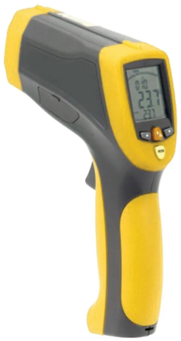
Fig. 1.1. A pyrometer, used for measuring temperature values for a specific area.

Fig. 1.2. An example of the thermographic measurement with a hand held camera.
1.3 Principles of Infrared Radiation
The origins of thermography can be traced back to the discovery of infrared radiation by the English astronomer Friedrich Wilhelm Herschel in 1800. Infrared radiation is the result of the movement of electrons, and is transmitted from the body surface as electromagnetic waves of varying wavelengths, ranging from 0.7 to 1000 μm (Fig. 1.3). Electromagnetic waves are produced across a spectrum of wavelengths, each wavelength representing a specific type of energy, such as X-rays, ultraviolet light, etc. (Fig. 1.3). A thermal camera measures energy in a specific part of the infrared waveband and correlates this with body surface temperature to produce a colour image illustrating the appropriate temperature values. In this way, body surface temperature is determined without physical contact with the examined animal (Kastberger and Stachl, 2003).

Fig. 1.3. The electromagnetic spectrum, with infrared radiation.
Part of the infrared band of the electromagnetic spectrum is absorbed into the atmosphere, which is why thermal equipment manufacturers produce thermographic cameras with only two types of sensor: short wave and long wave. Short-wave cameras typically detect radiation with wavelengths of 3–5 μm, whereas long-wave cameras are sensitive to wavelengths of 7–14 μm. According to Wien’s law (Kastberger and Stachl, 2003), an object with a high surface temperature emits peak radiation at short wavelengths (0.9–5 μm), whereas an object with a lower surface temperature emits peak radiation of long wavelengths (7–14 μm). Therefore, short-wave cameras are designed mostly for industrial use, where high temperature values are recorded, whereas long-wave cameras are used to read lower temperature values, e.g. in the civil engineering sector.
The animal body emits infrared radiation of wavelengths from around 3 to 50 μm. The peak emitted wavelength depends on the ambient temperature. This is related to the following rule: the higher the ambient temperature, the higher the body surface temperature, which results in radiation of shorter wavelengths being emitted. In the case of low ambient temperatures, the lower body surface temperature results in longer-wavelength radiation being emitted (Fig. 1.4). Due to the ambient temperature range typically encountered, long-wave thermal cameras are preferred for animal examinations.

Fig. 1.4. The warm right hand emits short-wave infrared radiation, while the cold left hand emits long-wave infrared radiation.
The body surface of an animal emits infrared electromagnetic waves, thus losing heat, but at the same time it also absorbs heat due to infrared radiation emitted or reflected from other sources, such as other animals, walls and the ground (Fig. 1.5). This can have a significant impact on the recorded body surface temperature (Cena, 1974). The ability of the body to absorb and reflect infrared radiation from other heat sources is a major reason for interference in body surface temperature measurements when examining animals and must be borne in mind when using thermography.

Fig. 1.5. The body surface of the horse emits infrared radiation, and also absorbs and reflects it from surrounding objects.
The emitted as well as the absorbed and reflected infrared radiation propagates isotropically (in all directions) from the entire body surface of the animal (Fig. 1.6).

Fig. 1.6. Isotropic emission of infrared radiation from a horse’s body surface.
1.4 The Thermographic Image
The measurement of electromagnetic wave energy emitted by the body using a thermal camera enables the creation of a thermographic image (thermogram). A thermogram is created by converting infrared radiation into electrical signals, which are then changed into visible-light radiation. This forms an image in a colour palette, in which various colours represent the relevant temperature range. This creates a graphical temperature distribution map of the examined body surface area. The colour bar appearing on the side of the thermogram maps each colour to a temperature range.
Thermograms presented in a simple ‘rainbow’ palette illustrate the warmest parts of the body in white and red, cooler areas in green and yellow, and the coolest parts in blue and black (Fig. 1.7). The thermograms in this book will be presented in this palette.
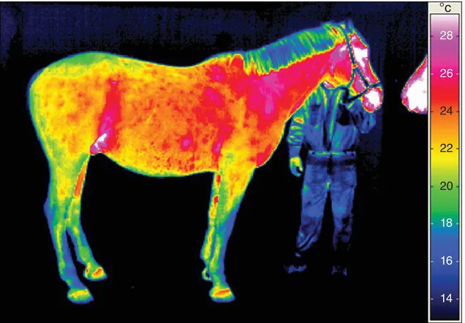
Fig. 1.7. Thermogram of the right lateral aspect of the horse. The warmest areas of the body are presented as white and red, yellow and green indicate cooler areas, and blue and black represent the coldest areas of the body.
An accurate measurement of an animal’s body surface temperature is dependent on the determination of the emissivity of the body surface. Emissivity is a measure of the efficiency of the emission of infrared radiation by any given body. Its value can range from 0 to 1, and depends on the emitting surface structure. Emissivity is low in the case of smooth surfaces but high for porous surfaces with a complex structure, such as skin (depending on the skin thickness and hair length). Biological tissues have emissivity values in the range of 0.95–0.98. To obtain the correct manual emissivity setting for a thermal camera, the following steps are carried out:
• Measure the temperature value of a given body area of the animal with a contact thermometer.
• Adjust the temperature reading on the display of the camera to the temperature reading on the contact thermometer.
In practice, emissivity is usually assumed to be 1, which will have a minimal impact on temperature accuracy.
1.5 Thermographic Imaging Technology
Thermographic camera technology has advanced rapidly in recent years, and a number of manufacturers now offer...
Table of contents
- Cover
- Half Title
- Title
- Copyright
- Contents
- About the Authors
- Acknowledgements
- Glossary
- Introduction
- 1 Principles of Equine Thermography
- 2 Fundamentals of Thermographic Examination
- 3 Interpretation of Thermographic Images and the Normal Superficial Temperature Distribution of the Horse
- 4 Development of Equine Thermography and its Use in Equestrianism
- 5 Use of Thermography in Physiotherapy
- 6 Recommendations for Thermography Application
- References
- Appendix 1: Equine Thermographic Examination Questionnaire
- Index
- Back Cover
Frequently asked questions
Yes, you can cancel anytime from the Subscription tab in your account settings on the Perlego website. Your subscription will stay active until the end of your current billing period. Learn how to cancel your subscription
No, books cannot be downloaded as external files, such as PDFs, for use outside of Perlego. However, you can download books within the Perlego app for offline reading on mobile or tablet. Learn how to download books offline
Perlego offers two plans: Essential and Complete
- Essential is ideal for learners and professionals who enjoy exploring a wide range of subjects. Access the Essential Library with 800,000+ trusted titles and best-sellers across business, personal growth, and the humanities. Includes unlimited reading time and Standard Read Aloud voice.
- Complete: Perfect for advanced learners and researchers needing full, unrestricted access. Unlock 1.5M+ books across hundreds of subjects, including academic and specialized titles. The Complete Plan also includes advanced features like Premium Read Aloud and Research Assistant.
We are an online textbook subscription service, where you can get access to an entire online library for less than the price of a single book per month. With over 1.5 million books across 990+ topics, we’ve got you covered! Learn about our mission
Look out for the read-aloud symbol on your next book to see if you can listen to it. The read-aloud tool reads text aloud for you, highlighting the text as it is being read. You can pause it, speed it up and slow it down. Learn more about Read Aloud
Yes! You can use the Perlego app on both iOS and Android devices to read anytime, anywhere — even offline. Perfect for commutes or when you’re on the go.
Please note we cannot support devices running on iOS 13 and Android 7 or earlier. Learn more about using the app
Please note we cannot support devices running on iOS 13 and Android 7 or earlier. Learn more about using the app
Yes, you can access Equine Thermography in Practice by Maria Soroko-Dubrovina, Mina C G Davies Morel in PDF and/or ePUB format, as well as other popular books in Medicine & Equine Veterinary Science. We have over 1.5 million books available in our catalogue for you to explore.