- Includes a brand new chapter on emergency morphology, designed to make the clinical significance and urgency of certain laboratory findings clear for biomedical scientists and to assist trainee haematologists in the recognition of major clinically important abnormalities
- Contains exceptional full colour images throughout
- Introduces important basic concepts of hematology, setting haematological findings in a clinical context
- Provides a fully updated self-assessment section
- An essential resource for trainee haematologists, biomedical scientists, and biomedical science and medical students
- English
- ePUB (mobile friendly)
- Available on iOS & Android
eBook - ePub
A Beginner's Guide to Blood Cells
About this book
The third edition of this popular pocket book, A Beginner's Guide to Blood Cells written by Professor Barbara Bain, provides a concise introduction to normal and abnormal blood cells and blood counts for trainees in haematology.
Trusted by 375,005 students
Access to over 1.5 million titles for a fair monthly price.
Study more efficiently using our study tools.
Information
CHAPTER 1
The Blood Film and Count
Blood
Blood is a life‐sustaining fluid that circulates through the heart and blood vessels. It carries oxygen and nutrients to the tissues and waste products to the lungs, liver and kidneys, where they can be removed from the body. Usually when blood is removed from the body it forms a solid blood clot. However, if clotting is prevented by mixing with an anticoagulant, the blood separates, under the influence of gravity, into three layers (Fig. 1.1). The bottom layer is deep red in colour and is composed of red cells. The top layer is clear and pale yellow. It is called plasma and is composed of various salts and proteins dissolved in water. In between is a narrow layer called the buffy coat because of its buff or yellowish white colour. The buffy coat is composed mainly of cells of a variety of types, collectively known as white cells. In addition there are small cellular fragments, called platelets, which have a role in blood clotting.
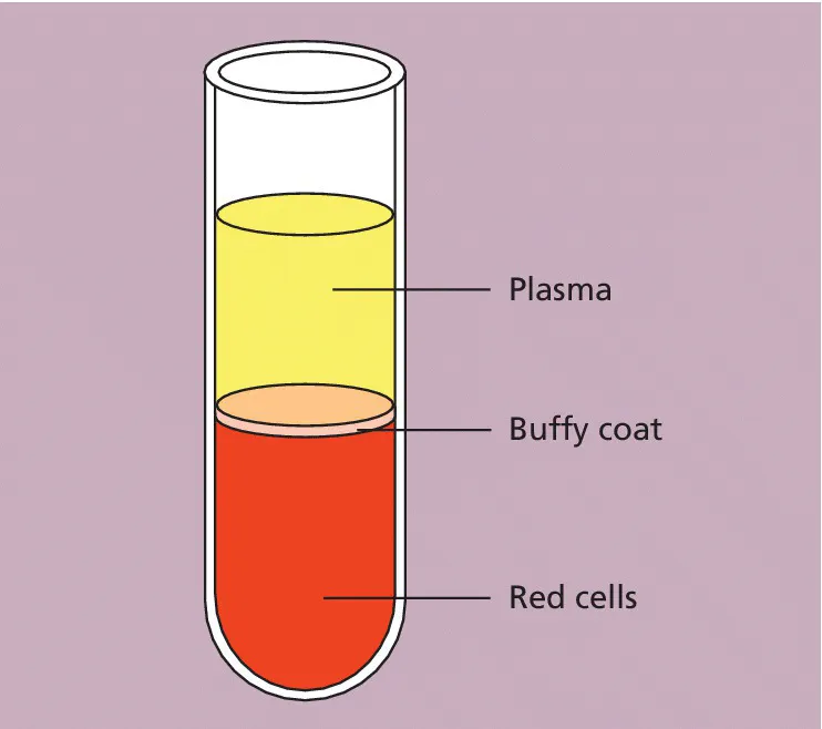
Fig. 1.1 Diagram of a tube of anticoagulated blood that has been allowed to sediment, showing the separation of blood into red cells, a buffy coat (white cells and platelets) and plasma.
The blood film
Although we can judge the proportions of red cells and white cells in a tube of sedimented blood, we get far more information if the blood is carefully mixed and a thin layer is spread on a glass slide to form a blood film. The blood cells are then preserved by exposure to the alcohol methanol, a process known as fixation. The fixed film of blood is stained with a mixture of several dyes so that the individual cells can be recognized when they are examined with a microscope. After staining, the colour of red cells is enhanced and the white cells and platelets, which would otherwise be transparent and colourless, have acquired a variety of colours that allow their detailed structure to be recognized. One of the commonest mixtures of dyes used to stain blood cells is the May–Grünwald–Giemsa (MGG) stain, named after its inventors. All the photographs in this book are of MGG‐stained blood films.
Red cells
The most numerous cells in a blood film are the red cells, also known as erythrocytes. Normal red cells are disc‐shaped but are thinner in the centre (Fig. 1.2). As a consequence, on a stained blood film, they have a circular outline and a paler central area (Fig. 1.3). Red cells owe their pinkish‐brown colour to the presence of a complex protein, haemoglobin, which is their major constituent. Enhancement of their colour in a stained film is because haemoglobin takes up eosin, one of the dyes of the MGG stain. In the body it is haemoglobin of the red cells that, in the lungs, combines with oxygen from inspired air and transports it to tissues where it is needed for the metabolic processes supplying the energy needs of the body. Mature red cells in humans (although not in some other species) differ from most body cells in that they do not have a nucleus. Red cells are produced in the bone marrow and usually lose their nuclei when they are released into the blood stream.

Fig. 1.2 A diagram of a red cell viewed from above and in cross‐section.
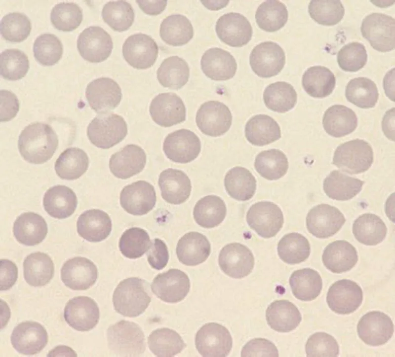
Fig. 1.3 Normal red cells (erythrocytes) showing little variation in size and shape, an approximately round outline and a small area of central pallor in some of the cells. The small structures containing lilac‐staining granules between the red cells are platelets.
White cells
In healthy people there are at least five types of white cell, or leucocyte, in the circulating blood. Unlike red cells, white cells have retained their nuclei. The cell is therefore made up of a nucleus and cytoplasm. The cytoplasm is the site of protein synthesis and other cellular functions. The nucleus is composed of chromatin, which is mainly deoxyribonucleic acid (DNA), carrying genetic messages. Genetic messages are transmit...
Table of contents
- Cover
- Title Page
- Table of Contents
- Preface
- Abbreviations
- CHAPTER 1: The Blood Film and Count
- CHAPTER 2: Assessing Red Cells
- CHAPTER 3: Assessing White Cells and Platelets
- CHAPTER 4: Haematological Findings in Health and Disease
- CHAPTER 5: Emergency Morphology: The Relevance of the Full Blood Count and Blood Film in Acute Illness
- CHAPTER 6: Self‐assessment
- Index
- End User License Agreement
Frequently asked questions
Yes, you can cancel anytime from the Subscription tab in your account settings on the Perlego website. Your subscription will stay active until the end of your current billing period. Learn how to cancel your subscription
No, books cannot be downloaded as external files, such as PDFs, for use outside of Perlego. However, you can download books within the Perlego app for offline reading on mobile or tablet. Learn how to download books offline
Perlego offers two plans: Essential and Complete
- Essential is ideal for learners and professionals who enjoy exploring a wide range of subjects. Access the Essential Library with 800,000+ trusted titles and best-sellers across business, personal growth, and the humanities. Includes unlimited reading time and Standard Read Aloud voice.
- Complete: Perfect for advanced learners and researchers needing full, unrestricted access. Unlock 1.5M+ books across hundreds of subjects, including academic and specialized titles. The Complete Plan also includes advanced features like Premium Read Aloud and Research Assistant.
We are an online textbook subscription service, where you can get access to an entire online library for less than the price of a single book per month. With over 1.5 million books across 990+ topics, we’ve got you covered! Learn about our mission
Look out for the read-aloud symbol on your next book to see if you can listen to it. The read-aloud tool reads text aloud for you, highlighting the text as it is being read. You can pause it, speed it up and slow it down. Learn more about Read Aloud
Yes! You can use the Perlego app on both iOS and Android devices to read anytime, anywhere — even offline. Perfect for commutes or when you’re on the go.
Please note we cannot support devices running on iOS 13 and Android 7 or earlier. Learn more about using the app
Please note we cannot support devices running on iOS 13 and Android 7 or earlier. Learn more about using the app
Yes, you can access A Beginner's Guide to Blood Cells by Barbara J. Bain in PDF and/or ePUB format, as well as other popular books in Medicine & Hematology. We have over 1.5 million books available in our catalogue for you to explore.