These volumes teach readers to think beyond apoptosis and describes all of the known processes that cells can undergo which result in cell death
This two-volume source on how cells dies is the first, comprehensive collection to cover all of the known processes that cells undergo when they die. It is also the only one of its kind to compare these processes. It seeks to enlighten those in the field about these many processes and to stimulate their thinking at looking at these pathways when their research system does not show signs of activation of the classic apoptotic pathway. In addition, it links activities like the molecular biology of one process (eg. Necrosis) to another process (eg. apoptosis) and contrasts those that are close to each.
Volume 1 of Apoptosis and Beyond: The Many Ways Cells Die begins with a general view of the cytoplasmic and nuclear features of apoptosis. It then goes on to offer chapters on targeting the cell death mechanism; microbial programmed cell death; autophagy; cell injury, adaptation, and necrosis; necroptosis; ferroptosis; anoikis; pyronecrosis; and more. Volume 2 covers such subjects as phenoptosis; pyroptosis; hematopoiesis and eryptosis; cyclophilin d-dependent necrosis; and the role of phospholipase in cell death.
Covers all known processes that dying cells undergo
Provides extensive coverage of a topic not fully covered before
Offers chapters written by top researchers in the field
Provides activities that link and contrast processes to each other
Apoptosis and Beyond: The Many Ways Cells Die will appeal to students and researchers/clinicians in cell biology, molecular biology, oncology, and tumor biology.
Trusted by 375,005 students
Access to over 1.5 million titles for a fair monthly price.
1 General View of the Cytoplasmic and Nuclear Features of Apoptosis
Humberto De Vitto,1 Juan P. Valencia,2 and James A. Radosevich3
1Center of Health and Science, Federal University of Rio de Janeiro, Rio de Janeiro, Brazil
2University of Rio de Janeiro, Rio de Janeiro, Brazil
3Department of Oral Medicine and Diagnostic Sciences, University of Illinois at Chicago, Chicago, IL, USA
Abbreviations
AIF
apoptosis-inducing factor
Apaf-1
apoptotic protease activating factor-1
ATP
adenosine triphosphate
BA
bongkrekic acid
Bcl-2
B-cell lymphoma-2
BID
BH3-interacting domain death agonist
bp
base pair
CAD
caspase-activated DNase
c-FLIP
cellular FLICE-inhibitory protein
Chx
cyclohexamide
CsA
cyclosporine A
CTL
cytotoxic T lymphocyte
cyt c
cytochrome c
DISC
death-inducing signal complex
DED
death effector domain
endo D
endonuclease D
endo G
endonuclease G
ER
endoplasmic reticulum
FADD
Fas-associated death-domain protein
FasL
fatty acid synthetase ligand
FasR
fatty acid synthetase receptor
Gzm-A
granzyme-A
Gzm-B
granzyme-B
ICAD
inhibitor of caspase-activated DNase
Kb
kilobase
MEF
mouse embryonic fibroblast
MOMP
mitochondrial outer-membrane permeabilization
NK
natural killer
OMM
outer mitochondrial membrane
PCD
programmed cell death
PFN
Perforin
PT
pore transition
RIP
receptor-interacting protein
ROCK I
Rho effector protein
ROS
reactive oxygen species
SET
stress-response complex
tBID
truncated BID
TNF
tumor necrosis factor
TNFR1
tumor necrosis factor receptor 1
TRADD
TNF receptor-associated death domain
TUNEL
terminal deoxynucleotide tranferase dUTP nick end labeling
1.1 Introduction
The normal development of a cell and the life cycles of the multicellular organism rely on a finely tuned balance between cell survival and death. In a biological context, cells need to grow, divide, and die. In regard to the latter process, cells have developed a very precisely regulated means of programmed cell death (PCD), which contributes to the maintenance of normal cell turnover, leading to reduced impact on tissues, organs, and the organism itself. Some cells have evolved a PCD process called apoptosis. Apoptosis can be simply defined as a set of biochemical cytoplasmic and mitochondrial events that may lead to the execution phase of nuclear events.
A wide array of stress stimuli can trigger the apoptotic process, and the biochemical signal can then be amplified in the cytoplasm and mitochondria by both extrinsic and intrinsic pathways. The convergence of the apoptotic signal is considered the activation of a family of cysteine aspartyl-specific proteases (caspases), composed of 12 proteins strictly involved in the apoptotic cell death process. The dying cells activate the execution pathway that leads to the appearance of blebs and to the “pinching off” of many of them, forming “apoptotic bodies,” which may be rounded and retracted from their own tissue. Subsequently, the immune system cells are able to eliminate the apoptotic bodies through an engulfment cell process. The morphological and biochemical features during the apoptotic process are not fully understood.
At the nuclear level, it is well established that endonucleases and exonucleases may hydrolyze the DNA into small fragments (200 pb) [1]. The nuclear events depend on caspase activation. Caspase 3 is considered the most important protease of the executioner pathway, and is activated by different initiator caspases. For instance, caspase 8 is activated from the death receptor, caspase 9 is involved in the mitochondrial apoptotic process, and caspase 10 is involved in the Perforin/granzyme (PFN/Gzm) pathways. The cleaved caspase 3 cleaves the endonuclease caspase-activated DNase (CAD), degrading the DNA at nucleosomal linkers [2,3], which generates small DNA fragments (∼50–300 kb). The subsequent processing of the DNA by exonucleases and endonucleases leads to the formation of 200 bp fragments. Many organelles, such as the Golgi apparatus, endoplasmic reticulum (ER), lysosomes, and mitochondria, can be recycled or eliminated, depending on the apoptotic stimuli. It is important to note that mitochondria play a pivotal role in apoptosis, since they can release cytochrome c (cyt c) and endonuclease D (endo D), leading to cell death [4,5].
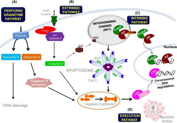
One of the apoptotic pathways is the extrinsic or death-receptor pathway. It depends for its activation on a death domain and a death ligand, such as tumor necrosis factor alpha (TNFα) and tumor necrosis factor receptor 1 (TNFR1). The ligand represents the external death signal, leading to the intracellular signaling of the effector pathway. The main receptors recruit adaptor proteins like Fas-associated death-domain protein (FADD), TNF receptor-associated death domain (TRADD), and receptor-interacting protein (RIP) [6–8], which in turn recruit other molecules such as pro-caspase 8. The dimerization of the death effector domain (DED) leads to the formation of a death-inducing signal complex (DISC), triggering the subsequent process of autocatalysis of pro-caspase 8 to an activated protein (caspase 8) [9]. Caspase 8 activation is considered the main feature that starts the extrinsic pathway, leading to cell death. In many cases, depending on the apoptotic stimuli, the extrinsic pathway can crosstalk with the intrinsic pathway through proteolysis of the BH3-only protein, BH3-interacting domain death agonist (BID), which is what promotes the release of cyt c from the mitochondria into the cytoplasm. In the cytoplasm, cyt c may be assembled with the adaptor protein apoptotic protease activating factor-1 (Apaf-1) and ATP, generating in the cytosol the multimolecular holoenzyme complex called the “apoptosome” (Figure 1.1) [10].
Figure 1.1 Schematic representation of the cytoplasmic and nuclear events of apoptosis. The Perforin/Granzyme pathway, extrinsic pathway, and intrinsic pathway represent the three main pathways of apoptosis. Through a vast array of death signals, all three pathways can be triggered. (A) The Perforin/Granzyme pathway is a unique pathway that partially works in a caspase-independent fashion (granzyme A branch), leading directly to DNA cleavage and cell death. However, the activation of the granzyme B branch can trigger initiator caspase 10, which activates executioner caspase 3. (B) The extrinsic pathway, when activated, can cleave pro-caspase 8 to caspase 8 by FAAD, then activate executioner caspase 3. Caspase 8 plays an important role in the activation of a truncated BID (tBID) protein, leading to the release of mitochondria proteins like cyt c. (C) Upon receiving incoming signals, the intrinsic pathway induces MPTP opening, leading to the release from the mitochondria of proteins such as cyt c, endo G/AIF, and Htra2/Omi. On the cytosol, cyt c forms the apoptosome, which cleaves pro-caspase ...
Table of contents
Cover
Title Page
Copyright
List of Contributors
Chapter 1: General View of the Cytoplasmic and Nuclear Features of Apoptosis
Chapter 2: Mitochondria in Focus: Targeting the Cell-Death Mechanism
Chapter 3: Microbial Programmed Cell Death
Chapter 4: Autophagy
Chapter 5: Cell Injury, Adaptation, and Necrosis
Chapter 6: Necroptosis
Chapter 7: Ferroptosis
Chapter 8: Anoikis Regulation: Complexities, Distinctions, and Cell Differentiation
Chapter 21: Alkylating-Agent Cytotoxicity Associated with O6-Methylguanine
Chapter 22: Entosis
Chapter 23: Mitotic Catastrophe
Chapter 24: NETosis and ETosis: Incompletely Understood Types of Granulocyte Death and their Proposed Adaptive Benefits and Costs
Chapter 25: Parthanatos: Poly ADP Ribose Polymerase (PARP)-Mediated Cell Death
Chapter 26: Methuosis: Drinking to Death
Chapter 27: Oncosis
Chapter 28: Autoschizis: A Mode of Cell Death of Cancer Cells Induced by a Prooxidant Treatment In Vitro and In Vivo
Chapter 29: Programmed Death 1 (PD1)-Mediated T-Cell Apoptosis and Cancer Immunotherapy
Index
End User License Agreement
Frequently asked questions
Yes, you can cancel anytime from the Subscription tab in your account settings on the Perlego website. Your subscription will stay active until the end of your current billing period. Learn how to cancel your subscription
No, books cannot be downloaded as external files, such as PDFs, for use outside of Perlego. However, you can download books within the Perlego app for offline reading on mobile or tablet. Learn how to download books offline
Perlego offers two plans: Essential and Complete
Essential is ideal for learners and professionals who enjoy exploring a wide range of subjects. Access the Essential Library with 800,000+ trusted titles and best-sellers across business, personal growth, and the humanities. Includes unlimited reading time and Standard Read Aloud voice.
Complete: Perfect for advanced learners and researchers needing full, unrestricted access. Unlock 1.5M+ books across hundreds of subjects, including academic and specialized titles. The Complete Plan also includes advanced features like Premium Read Aloud and Research Assistant.
Both plans are available with monthly, semester, or annual billing cycles.
We are an online textbook subscription service, where you can get access to an entire online library for less than the price of a single book per month. With over 1.5 million books across 990+ topics, we’ve got you covered! Learn about our mission
Look out for the read-aloud symbol on your next book to see if you can listen to it. The read-aloud tool reads text aloud for you, highlighting the text as it is being read. You can pause it, speed it up and slow it down. Learn more about Read Aloud
Yes! You can use the Perlego app on both iOS and Android devices to read anytime, anywhere — even offline. Perfect for commutes or when you’re on the go. Please note we cannot support devices running on iOS 13 and Android 7 or earlier. Learn more about using the app
Yes, you can access Apoptosis and Beyond by James A. Radosevich in PDF and/or ePUB format, as well as other popular books in Ciencias biológicas & Biología celular. We have over 1.5 million books available in our catalogue for you to explore.