Pathology of Liver Diseases
eBook - ePub

Pathology of Liver Diseases

  1. English
  2. ePUB (mobile friendly)
  3. Available on iOS & Android
eBook - ePub

Pathology of Liver Diseases

About this book

Pathology of Liver Diseases is a rapid reference consultation tool that uses both book and online material to present a whole range of liver disorders. The book emphasizes not only the pathology seen in biopsy and surgical material, but also the most pertinent clinical and laboratory findings including epidemiology, etiologic and pathophysiologic concepts, and the differential diagnoses. Key references appear at the end of each chapter.

The book is also accompanied by a companion website: www.wiley.com/go/kanel/liverpathology

It contains the following online material:

  • A complete Reference List.
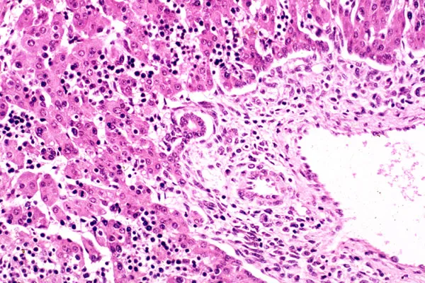
  • A Library that contains over 860 images of the various liver diseases, which adds to over 540 images that are in the book itself
  • Additional Tables that address in detail the grading and staging of various liver diseases such as viral
    hepatitis and fatty liver diseases.
  • 140 Case Examples, which include over 420 images that demonstrate the various ways many of these disease entities clinically present.
  • A PowerPoint presentation entitled "Liver Transplantation โ€“ Surgical Procedure", which includes photographs from the operating table of the step-by-step process in liver transplantation.

Pathology of Liver Diseases provides gastroenterologists and pathologists with a multi-media, well-illustrated, and concise guide to the pathology and clinical diagnoses of liver disorders.

Frequently asked questions

Yes, you can cancel anytime from the Subscription tab in your account settings on the Perlego website. Your subscription will stay active until the end of your current billing period. Learn how to cancel your subscription.
No, books cannot be downloaded as external files, such as PDFs, for use outside of Perlego. However, you can download books within the Perlego app for offline reading on mobile or tablet. Learn more here.
Perlego offers two plans: Essential and Complete
  • Essential is ideal for learners and professionals who enjoy exploring a wide range of subjects. Access the Essential Library with 800,000+ trusted titles and best-sellers across business, personal growth, and the humanities. Includes unlimited reading time and Standard Read Aloud voice.
  • Complete: Perfect for advanced learners and researchers needing full, unrestricted access. Unlock 1.4M+ books across hundreds of subjects, including academic and specialized titles. The Complete Plan also includes advanced features like Premium Read Aloud and Research Assistant.
Both plans are available with monthly, semester, or annual billing cycles.
We are an online textbook subscription service, where you can get access to an entire online library for less than the price of a single book per month. With over 1 million books across 1000+ topics, weโ€™ve got you covered! Learn more here.
Look out for the read-aloud symbol on your next book to see if you can listen to it. The read-aloud tool reads text aloud for you, highlighting the text as it is being read. You can pause it, speed it up and slow it down. Learn more here.
Yes! You can use the Perlego app on both iOS or Android devices to read anytime, anywhere โ€” even offline. Perfect for commutes or when youโ€™re on the go.
Please note we cannot support devices running on iOS 13 and Android 7 or earlier. Learn more about using the app.
Yes, you can access Pathology of Liver Diseases by Gary C. Kanel in PDF and/or ePUB format, as well as other popular books in Medicine & Pathology. We have over one million books available in our catalogue for you to explore.

Information

Year
2017
Print ISBN
9781118895030
eBook ISBN
9781118895009
Edition
1
Subtopic
Pathology

1
Normal Liver

The liver is a unique organ that has numerous structural and physiological functions. It is most important when discussing liver pathology that one understands first the normal liver histology before one can best understand the basic pathophysiologic concepts of the numerous liver diseases. The pathologist plays a fundamental role in assessing the various morphologic features seen in liver tissue, whether by fine needle aspirates, needle or wedge biopsies, partial hepatectomies, liver explants, or autopsy material. The pathologist also has not only routine but also numerous special histochemical and immunohistologic stains as well. Yet correlating the histologic findings with the most pertinent clinical and laboratory data enables the pathologist to better arrive at a diagnosis and the most pertinent differential possibilities.
This introductory chapter addresses all aspects of the normal liver, reviewing the embryologic development, gross and microscopic features, the pertinent intracytoplasmic components and how their function varies with their location within the hepatic lobule, and the importance of stem cell function within the liver. Additionally the various useful stains and laboratory values will also be presented, as well as a brief outline of how best to organize pathologic readings and signouts of liver biopsy specimens.

Embryology

The hepatic primordium anlage initially appears at the end of the third week of gestation and is first seen as a hollow midline outgrowth stalk (hepatic diverticulum) of the endodermal epithelium at the distal aspect of the foregut. By the fourth week, the diverticulum enlarges from proliferation of the endodermal cell strands (hepatoblasts) and projects cranially into the mesoderm of the septum transversum, eventually giving rise to the liver hepatic parenchyma and intrahepatic ducts. The cephalic end ultimately develops into the right and left hepatic lobes, while the stalk between the diverticulum and foregut narrows and forms the extrahepatic biliary system and gallbladder.
Solid cords are initially formed by proliferating endodermal cells. These eventually anastomose to form vesicles and cribriform tubules with centrally located lumenal structures (biliary canaliculi). The cords eventually merge and develop small channels and capillaries that subdivide the cords to eventually form the hepatic sinusoids. The individual hepatoblasts are progenitor cells that develop into mature hepatocytes, with those immediately adjacent to the portal mesenchyme becoming the ductal plates. The rapid growth rate of the hepatic cords enables the development of sheets of cells (muralium multiplex) that persist until birth, after which the cell sheets narrow to two cells (muralium duplex) and eventually evolve within the first year of life into a one cell thick trabecular cord (muralium simplex). The perisinusoidal cells and Kupffer cells appear by three months gestation.
The mesoderm from the septum transversum initially surrounds the liver and is directly in contact with the lesser curvature of the stomach, duodenum, and ventral body wall. The mesoderm eventually forms the lesser omentum, the falciform, coronary, and triangular ligaments, with a portion developing into the liver (Glisson) capsule. The mesoderm on the liver surface is also in continuity with the peritoneum, and the portion that makes contact with the future diaphragm remains uncovered (bare area). The developing hepatic artery and vagus nerve branches follow the mesoderm along and adjacent to the portal vein.
The mesoderm is the main focus in the development of hematopoiesis, which begins at about 6 weeks and becomes most active during the fifth month of gestation. This process regresses with increase in bone marrow activity. The erythroid precursors are most prominent during fetal development within the hepatic sinusoids while the myeloid and megakaryocytic precursors reside mostly within the portal structures (Figure 1.1). This hematopoiesis is responsible for the enlarged size of the liver (up to 10% body weight by the tenth week of gestation, with the right and left lobes taking up an equal volume), but this size significantly regresses at birth (5% of body weight) at which time only rare small clusters of normoblasts can be seen. By 4 weeks of age hematopoietic activity has usually ceased. Additionally with time the left lobe diminishes in size, and the caudate and quadrate lobes develop as subdivisions of the right lobe.
Image described by caption and surrounding text.
Figure 1.1 Embryonic development. A developing bile ductule is seen at the border of the portal tract and parenchyma. The portal tract and sinusoids contain hematopoietic precursors (extramedullary hematopoiesis).
The vascular network, originally derived from the development of ...

Table of contents

  1. Cover
  2. Title Page
  3. Copyright
  4. Preface
  5. About the companion website
  6. 1: Normal Liver
  7. 2: Viral Hepatitis
  8. 3: Fatty Liver Diseases
  9. 4: Diseases of the Biliary Tract
  10. 5: Non-Viral Infectious Diseases
  11. 6: Granulomatous Hepatitis
  12. 7: Autoimmune Hepatitis
  13. 8: Vascular Disorders
  14. 9: Genetic and Metabolic Hepatic Diseases
  15. 10: Developmental Hepatobiliary Disorders and Cystic Diseases
  16. 11: Drug- and Toxin-Induced Liver Diseases
  17. 12: Liver Transplantation
  18. 13: Hepatic Tumors, Benign
  19. 14: Hepatic Tumors, Malignant
  20. 15: Miscellaneous Hepatic Disorders
  21. Index
  22. EULA