Guide to Canine and Feline Electrocardiography
eBook - ePub

Guide to Canine and Feline Electrocardiography

  1. English
  2. ePUB (mobile friendly)
  3. Available on iOS & Android
eBook - ePub

About this book

Guide to Canine and Feline Electrocardiography offers a comprehensive and readable guide to the diagnosis and treatment of abnormal heart rhythms in cats and dogs.

  • Covers all aspects of electrocardiography, from basics to advanced concepts of interest to specialists
  • Explains how to obtain high-quality electrocardiograms
  • Offers expert insight and guidance on the diagnosis and treatment of simple and complex arrhythmias alike
  • Features numerous case examples, with electrocardiograms and Holter monitor recordings
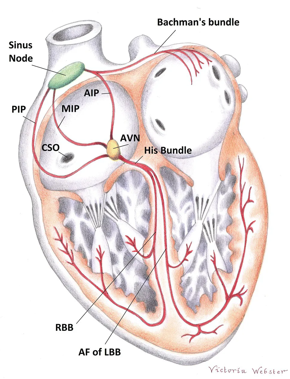
  • Shows the characteristics of normal and abnormal heart rhythms in dogs and cats
  • Includes access to a website with self-assessment questions and the appendices and figures from the book

Frequently asked questions

Yes, you can cancel anytime from the Subscription tab in your account settings on the Perlego website. Your subscription will stay active until the end of your current billing period. Learn how to cancel your subscription.
At the moment all of our mobile-responsive ePub books are available to download via the app. Most of our PDFs are also available to download and we're working on making the final remaining ones downloadable now. Learn more here.
Perlego offers two plans: Essential and Complete
  • Essential is ideal for learners and professionals who enjoy exploring a wide range of subjects. Access the Essential Library with 800,000+ trusted titles and best-sellers across business, personal growth, and the humanities. Includes unlimited reading time and Standard Read Aloud voice.
  • Complete: Perfect for advanced learners and researchers needing full, unrestricted access. Unlock 1.4M+ books across hundreds of subjects, including academic and specialized titles. The Complete Plan also includes advanced features like Premium Read Aloud and Research Assistant.
Both plans are available with monthly, semester, or annual billing cycles.
We are an online textbook subscription service, where you can get access to an entire online library for less than the price of a single book per month. With over 1 million books across 1000+ topics, we’ve got you covered! Learn more here.
Look out for the read-aloud symbol on your next book to see if you can listen to it. The read-aloud tool reads text aloud for you, highlighting the text as it is being read. You can pause it, speed it up and slow it down. Learn more here.
Yes! You can use the Perlego app on both iOS or Android devices to read anytime, anywhere — even offline. Perfect for commutes or when you’re on the go.
Please note we cannot support devices running on iOS 13 and Android 7 or earlier. Learn more about using the app.
Yes, you can access Guide to Canine and Feline Electrocardiography by Ruth Willis, Pedro Oliveira, Antonia Mavropoulou, Ruth Willis,Pedro Oliveira,Antonia Mavropoulou in PDF and/or ePUB format, as well as other popular books in Medicine & Veterinary Medicine. We have over one million books available in our catalogue for you to explore.

Information

Year
2018
Print ISBN
9781119253846
eBook ISBN
9781119254317
Edition
1

1
Anatomy of the Conduction System

Pedro Oliveira

Introduction

The heart possesses a specialised conduction system that is responsible for generating and transmitting electrical stimuli to the whole heart in a specific and ordered fashion. It is composed of the sinoatrial node (SA), internodal and inter‐atrial pathways, atrioventricular junction, bundle branches and Purkinje fibres (Figure 1.1). The SA contains specialised ‘pacemaker’ cells that have the ability to spontaneously depolarise, generating electrical impulses. The remainder of the conduction system is composed mainly of cells organised in bundles that allow conduction of the electrical stimuli. These structures are present in the walls of the heart and are interwoven with the myocardial tissue itself. It is not possible to distinguish them from the rest of the myocardium (working myocardium) with the naked eye, only with certain stains under the microscope.
Cross section of the heart with lines pointing at the sinus node, Bachman's bundle, His bundle, AVN, RBB, AF of LBB, CSO, MIP, AIP, and PIP.
Figure 1.1 Cardiac conduction system. The sinoatrial node, also known as the sinus node, is found in the wall of the right atrium at its junction with the cranial vena cava in the upper portion of the terminal groove (sulcus terminalis). It is connected to the atrioventricular node (AVN) located on the floor of the right atrium via the anterior (AIP), middle (MIP) and posterior (PIP) internodal pathways. Connections also exist between the right and left atria, of which Bachmann’s bundle and the inferior interatrial pathway (not illustrated) are the most important. Bachmann’s bundle runs in the upper portion of the atrial septum towards the left auricle. The inferior interatrial pathway is composed of fibres that are continuous with the right atrial myocardium at the level of the ostium of the coronary sinus (CSO) and with the left atrial myocardium from which they separate approximately 20–30 mm from the CSO. The AVN is continued by the Bundle of His that penetrates the fibrous skeleton of the heart and ramifies into the right (RBB) and left (LBB) bundle branches. The LBB is composed of anterior (AF) and posterior ramifications (not illustrated). The bundle branch subdivisions give rise to numerous small branches that spread all over the subendocardium of both ventricles, forming the Purkinje network that connects the conduction system to the working myocardium.
The anatomy of these structures is presented in this chapter. To avoid confusion, the use of human anatomical terminology is avoided, and terminology commonly used in veterinary medicine for quadruped patients is preferred. Given the different orientation of the heart within the chest of dogs and cats in comparison to humans, the following terms are used: cranial instead of anterior; caudal instead of posterior; dorsal instead of superior; and ventral instead of inferior. However, since some terms are so widespread in veterinary literature (e.g. left anterior or posterior fascicles), it is difficult to avoid the use of such terms even though they are not entirely appropriate.

Sinoatrial node

Anatomy

The SA, also known as the sinus node, is found in the wall of the right atrium at its junction with the cranial vena cava in the upper portion of the terminal groove (sulcus terminalis) (Figure 1.1).
In dogs, it lies less than 1 mm beneath the epicardium and occupies almost the entire thickness of the atrial wall from epicardium to endocardium.1 The total size of the canine node was described as being approximately 5 mm3 with an oblong shape, although with significant variation observed amongst individuals.1,2 Other reports suggest a more extensive location of up to 3–4 cm between both venae cavae.3,4
In cats, according to one study including five male and five female domestic shorthair cats, the SA node was located 0.06–0.11 mm beneath the epicardium with an almost triangular shape.5 In males the reported size was 2.78 × 2.80 × 0.54 mm, and in females it was 2.75 × 2.64 × 0.45 mm.5 A different study involving 12 cats produced different results.6 A reconstruction of the SA node based on histological and electrophysiological data was performed in five of these cats, revealing an ellipsoid shape with a total area of 10.5 ± 0.76 mm2, a maximum length of 7.4 ± 0.74 mm, a maximal width of 2.2 ± 0.10 mm and a thickness of 0.41 ± 0.060 mm.6

Histology

Histologically, the SA is composed of specialised muscle fibres arranged in a network.7 Many small bundles are present with irregular courses interspersed with connective tissue accompanied by capillary vessels and nerve cells. The surface of the SA is covered by epicardium, and the remaining areas are surrounded by atrial muscle. Each nodal fibre shows a smooth transition to ordinary atrial muscle fibres at the periphery of the SA.
Three different types of cells are present: normal working myocardial cells, transitional cells and P (pacemaker) cells.
The P cells are responsible for the ability of the SA to spontaneously generate electrical stimuli. They represent approximately 50% of the cells in the SA and are also present in other areas of the conduction system (e.g. atrioventricular node [AVN]) in fewer numbers. They are organised in small groups of approximately five cells surrounded by connective tissue that function as a unit.
The transitional cells are also present in other parts of the conduction system (e.g. internodal tracts and the atrioventricular junction) and seem to provide a link between the specialised cells and the normal working myocardium.

Sinoatrial Exit Pathways

The existence of discrete exit sites has been described at the cranial and caudal ends of the canine sinus node.8 Ablation of these sites resulted in sinoatrial block, suggesting that the SA was not anatomically continuous with the atrial myocardium.8,9 It was suggested that vessels and connective tissue around the SA tissue were responsible for anatomical and physiological blocks on both sides of the node with the exception of the exit sites.9 These findings, together with the reports of a length of up to 3–4 cm,3,4 provide...

Table of contents

  1. Cover
  2. Table of Contents
  3. Preface
  4. 1 Anatomy of the Conduction System
  5. 2 Cardiac electrophysiology
  6. 3 Cardiac vectors and the genesis of the electrocardiogram
  7. 4 Electrocardiography
  8. 5 Sinus rhythms
  9. 6 Pathogenesis and classification of arrhythmias
  10. 7 Bradyarrhythmias and Conduction Disturbances
  11. 8 Atrial Rhythms
  12. 9 Atrial Fibrillation
  13. 10 Junctional Rhythms
  14. 11 Ventricular Rhythms
  15. 12 Clinical Approach to Arrhythmias and Intermittent Collapse
  16. 13 Diagnostic Approach to Narrow‐QRS Complex Tachycardia
  17. 14 Diagnostic Approach to Wide‐QRS Complex Tachycardia
  18. 15 Ambulatory Electrocardiographic Recordings
  19. 16 Heart Rate Variability
  20. 17 Anti‐arrhythmic Drugs
  21. 18 Pacemaker Therapy
  22. 19 Electrophysiology Studies and Catheter Ablation
  23. 20 Arrhythmias in Canine Cardiomyopathies and Valvular Heart Disease
  24. 21 Arrhythmias in Feline Cardiomyopathies
  25. 22 Inherited Ventricular Arrhythmias in German Shepherd Dogs
  26. 23 Systemic Disease and Arrhythmias, Including Selected Non‐cardiogenic Causes of Collapse
  27. 24 Cardiac Arrhythmias and Anaesthesia
  28. Appendix 1: Normal Ecg Measurements for Cats and Dogs
  29. Appendix 2: Arrhythmias – A Brief Review
  30. Appendix 3: Mean Electrical Axis
  31. Appendix 4: Anti‐Arrhythmic Drugs and Dosages*
  32. Appendix 5: Sample ECG Reports
  33. Self‐assessment
  34. Index
  35. End User License Agreement