Understanding Light Microscopy
eBook - ePub

Understanding Light Microscopy

Jeremy Sanderson

Share book
  1. English
  2. ePUB (mobile friendly)
  3. Available on iOS & Android
eBook - ePub

Understanding Light Microscopy

Jeremy Sanderson

Book details
Book preview
Table of contents
Citations

About This Book

Introduces readers to the enlightening world of the modern light microscope

There have been rapid advances in science and technology over the last decade, and the light microscope, together with the information that it gives about the image, has changed too. Yet the fundamental principles of setting up and using a microscope rests upon unchanging physical principles that have been understood for years. This informative, practical, full-colour guide fills the gap between specialised edited texts on detailed research topics, and introductory books, which concentrate on an optical approach to the light microscope. It also provides comprehensive coverage of confocal microscopy, which has revolutionised light microscopy over the last few decades.

Written to help the reader understand, set up, and use the often very expensive and complex modern research light microscope properly, Understanding Light Microscopy keeps mathematical formulae to a minimum—containing and explaining them within boxes in the text. Chapters provide in-depth coverage of basic microscope optics and design; ergonomics; illumination; diffraction and image formation; reflected-light, polarised-light, and fluorescence microscopy; deconvolution; TIRF microscopy; FRAP & FRET; super-resolution techniques; biological and materials specimen preparation; and more.

  • Gives a didactic introduction to the light microscope
  • Encourages readers to use advanced fluorescence and confocal microscopes within a research institute or core microscopy facility
  • Features full-colour illustrations and workable practical protocols

Understanding Light Microscopy is intended for any scientist who wishes to understand and use a modern light microscope. It is also ideal as supporting material for a formal taught course, or for individual students to learn the key aspects of light microscopy through their own study.

Frequently asked questions

How do I cancel my subscription?
Simply head over to the account section in settings and click on “Cancel Subscription” - it’s as simple as that. After you cancel, your membership will stay active for the remainder of the time you’ve paid for. Learn more here.
Can/how do I download books?
At the moment all of our mobile-responsive ePub books are available to download via the app. Most of our PDFs are also available to download and we're working on making the final remaining ones downloadable now. Learn more here.
What is the difference between the pricing plans?
Both plans give you full access to the library and all of Perlego’s features. The only differences are the price and subscription period: With the annual plan you’ll save around 30% compared to 12 months on the monthly plan.
What is Perlego?
We are an online textbook subscription service, where you can get access to an entire online library for less than the price of a single book per month. With over 1 million books across 1000+ topics, we’ve got you covered! Learn more here.
Do you support text-to-speech?
Look out for the read-aloud symbol on your next book to see if you can listen to it. The read-aloud tool reads text aloud for you, highlighting the text as it is being read. You can pause it, speed it up and slow it down. Learn more here.
Is Understanding Light Microscopy an online PDF/ePUB?
Yes, you can access Understanding Light Microscopy by Jeremy Sanderson in PDF and/or ePUB format, as well as other popular books in Physical Sciences & Science History & Practice. We have over one million books available in our catalogue for you to explore.

Information

Publisher
Wiley
Year
2019
ISBN
9781118696811

1
Our Eyes and the Microscope

Outline

1.1 Introduction
  • The eye-brain system
  • The features of the eye
1.2 How Our Eyes Work
  • Response to visible electromagnetic radiation
  • Rods and cones
  • Basis of colour vision
  • Eye sensitivity to brightness
  • Dark vision
  • Ability to see shades of grey
1.3 The Anatomy of the Eye
  • Anatomy of the eye
  • Floaters
  • Lens focusing
  • Macula and fovea
  • Visual acuity
  • Eye dominance
1.4 Aberrations of the Eye
  • Spherical aberration
  • Chromatic aberration
  • Astigmatism
  • Saccades – tiling our visual field
1.5 Binocular and Stereoscopic Vision
  • Stereoscopic vision
  • Parallax
1.6 Why We Need Optical Aids
  • Near point
  • Accommodation
  • Nearest distance of distinct vision
  • The magnifying glass
  • Magnification of a simple lens
1.7 Using Lenses to Correct Eye Defects
  • Myopia
  • Emetropia
  • Hyperopia
  • Presbyopia
1.8 Seeing the Scientific Image
  • Filling in and bias
  • Images as scientific data sets
1.9 Sizes of Objects
  • The requirement for measurement
  • BioNumbers database
1.10 Nomenclature and History
  • Nomenclature
1.11 Chapter Summary
  • 8 points
Key Reading
References
Further Reading
Internet Resources

1.1 Introduction

‘Ah, I see!’ demonstrates how dependent we are on our sight, and how it is linked inextricably with understanding. We are a visually oriented species, and our two eyes are our ‘window on the world’. It is easy to forget that not all animals depend upon sight to the extent that humans do. Bats use echolocating sonar to navigate, and bees exploit ultraviolet (UV) light to discriminate foliage. At the other end of the visible spectrum, vipers sense infrared radiation to detect warm blooded prey. For those creatures whose habitat is underground (e.g. moles), vision necessarily cedes priority to other senses such as touch and smell. Other animals, chiefly birds, are more dependent upon their sight than humans, with a much greater proportion of their head devoted to large eyes.
Via the optic nerve, the eyes are a direct extension of the brain. The human eye is a wonderful device. If it were a camera, it might boast autofocus, wide-angle lens, auto-exposure, high sensitivity to light, automatic colour balancing, one-hundred megapixel resolution (each retina contains around 100 million sensitive cells) and 3D imaging (when used in pairs). We can see in bright daylight or discriminate a single light in the dark several miles away. The eye has naturally evolved to suit its principal function: helping its owner navigate the world.
Many modern microscopes are designed to be operated from a computer screen, rather than be used by viewing the image directly down the eyepieces. Nevertheless, all images must eventually be seen by our eyes, so in this chapter we shall discuss how our eyes function. We shall consider how well our eyes are adapted for their purpose but also the limitations and aberrations that they suffer from. These limitations are relevant to microscopy, affecting how we acquire and interpret scientific images (i.e. data) using the light microscope. We shall see how a magnifying glass works and why the microscope was developed to assist our vision.

1.2 How Our Eyes Work

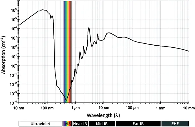
Any image must ultimately be seen by the eye. Light is electromagnetic radiation that stimulates the eye. It is merely a fraction of the entire electromagnetic spectrum (Figure 1.1). Only a small proportion of solar radiation reaches the earth’s surface. We depend on the ozone layer for protection; atmospheric dust, smoke, air molecules and water vapour also absorb a significant proportion of insolation, or incident solar radiation. Our eyes evolved from aquatic animals and contain a significant amount of water. Human sensitivity to electromagnetic radiation (Figure 1.2) corresponds closely to the wavelengths of minimum water absorbance, located away from harmful UV radiation and towards the infrared end of the spectrum.
Image is of the electromagnetic spectrum. It consists of two separate bands. The lower band represents wavelength (UV, visible, IR) that extends from 10 nm to 40000 nm. While the upper one represents the rays (Gamma, X-rays, UV, IR, and radio waves) that extends from 0.0001 nm to 100 m
Figure 1.1 The Electromagnetic spectrum The electromagnetic spectrum extends from beyond low frequencies used for modern radio to very high-frequency gamma radiation at the short-wavelength end of the spectrum, encompassing wavelengths from thousands of kilometres down to a fraction of a nanometre. Humans are able to discriminate the visible wavelengths from long wavelength, low energy, red at around 710 nm to short wavelength, high-energy violet radiation at around 380 nm. A very small number of people can see into the infrared, and people suffering from cataracts who have their lens removed (i.e. who are aphakic) can see into the UV, even if they can’t focus well.
Source: Glencross et al. 2011. Reproduced with permission from Oxford University Press.
Image shows a graph representing Human sensitivity to light. X-axis represents the wavelength and Y-axis the absorption. Different wavelengths represents different regions (visible, IR, vis).
Figure 1.2 Human sensitivity to light Visible light corresponds closely to the wavelengths of minimum water absorbance on the electromagnetic spectrum. See also Chapter 2, Section 2.5. Since our eyes are composed largely of water, it is advisable to use a heat filter (e.g. Schott KG5) in the light path to cut out any absorption of infrared light.
Source: Adapted from Kebes at English Wikipedia under the terms of the Creative Commons Attribution licence, CC BY-SA 3.0 (http://creativecommons.org/licenses/by-sa/3.0).
Our eyes respond to the visible part of the electromagnetic spectrum from near UV at a wavelength of 380 nm to deep red at 710 nm. This stimulation depends on both the energy (frequency, expressed as wavelength) and the quantity (number of photons) of light. The wavelength of light is perceived as colour, and the quantity of light (expressed as the amplitude of the light wave) is seen as intensity1 (see also Appendix A1.1.3 and Table A1.1, pages 45–46 in: Tilley, 2010). Suppose we have four LEDs: blue emitting at 490 nm; green at 555 nm; far red at 670 nm and infrared at 940 nm (commonly used for remote controls) all emitting the same radiant flux of 5 mW absolute power, measured in radiometric units. If we measure the respective light output of each of these LEDs in photometric units, the green LED will be the brightest (3.4 lm); the blue will be the second one (0.75 lm), and the far red will be the third (0.1 lm). The infrared LED will have a recorded emission of zero lumens (this example is taken from Tilley, 2010).
Further details about the electromagnetic spectrum and the nature of light are discussed in Chapter 2, Section 2.2 onwards. Light itself has no inherent colour; our perception of different hues is fundamentally a complex judgment experienced as a sensation by our brains. We discriminate colour very well, although brightness less so. This is why we choose paint in a range of colours, rather than different intensities.
The retina is the photo-sensitive tissue of the eye (Figure 1.3), withi...

Table of contents