Digital Planning and Custom Orthodontic Treatment offers a thorough overview of digital treatment planning as it relates to custom orthodontic treatment.
Covers 3D imaging of the dentition and the face with intraoral scanners, CBCT machines, and 3D facial scanners
Provides a complete guide to using digital treatment planning to improve the predictability, efficiency, and efficacy of orthodontic treatment
Discusses CAD/CAM fabrication of appliances and the monitoring of treatment progress and stability
Offers detailed descriptions for the main commercial systems on the market
Presents clinically oriented information to aid in yielding high quality and stable results
Trusted by 375,005 students
Access to over 1.5 million titles for a fair monthly price.
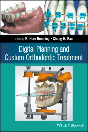
There is a growing demand for innovative ways to record the dentition and craniofacial complex. New technologies rely heavily on sophisticated tools and software to accurately capture the dentition. However, in order that these technologies are routinely used in a mainstream practice, a completely digital, highly accurate, and easily portable system needs to be established to create a worldwide information portal. A complete digital workflow will ensure that the appliances delivered are accurate and will be delivered efficiently to the consumer all over the world. Another more important benefit is that if appliances could be digitally built it would promise to reduce cost as the process would require less manual processing, and the transportation time and costs would not delay the fabrication process. It is this drive to create custom lab work that is fueling the next big game changer in orthodontics. The accurate representation of the dentition is by far the most important step to successful orthodontics. Traditional plaster casts are now slowly being replaced by digital models in orthodontics [1]. These digital models are often obtained via an indirect method that requires the transport of plaster casts or impressions of the dentition to a specialized company for laser or computer tomography (CT) scanning [2–5]. It is a known fact that the process of making plaster casts from dental impression materials such as alginate and polyvinyl siloxane (PVS) impression material has always some degree of dimensional change. During transportation and the period between the impression procedure and the pouring of plaster in the impression, the dimensions of the impression and thus the accuracy of the plaster model can change. Impressions have to be sterilized, transported to a dental lab, and, after fabrication of the plaster models, transported again to the orthodontic office. Plaster dental models have then to be stored in the dental office and retrieved during orthodontic treatment and are prone to fracture. Plaster models and also dental impressions can be scanned with desktop scanners with laser beams and with dedicated CT scanners to transform these models and impressions into digital dental models (Figure 1.1).
Figure 1.1 A dental model scanner. Company: 3Shape.
In the literature it is reported that the accuracy of digital dental models scanned directly from impressions when compared to the “golden standard, the plaster cast” is sufficient for orthodontic analysis and treatment planning. But for this method to get a digital dental model an impression or a plaster model is required. As the impression taking procedure and the fabrication of plaster casts is an indirect method to get digital dental models, there is interest in the use of a direct method to copy the dentition.
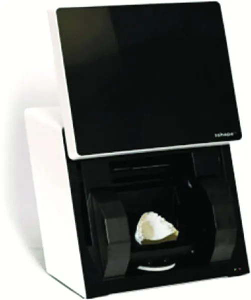
A direct method to capture the dentition is by using the cone beam computer tomography (CBCT) radiographs [6]. These radiographs can be used for dental analysis, but CBCT involves exposing the patient to radiation, and the quality of the dentition on the CBCT radiograph is directly related to the radiation dose used (Figure 1.2). Because of the ALARA principle (a radiation dose As Low As Reasonably Achievable), CBCT is not indicated for imaging of the dentition only.
Figure 1.2 The segmented dentition on a CBCT radiograph. Company: Anatomage Inc.
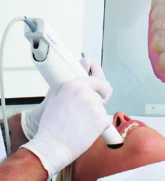
To answer the need for a digital yet economical solution to physical impression materials, several companies have developed intraoral scanning systems to acquire digital intraoral impressions for any type of dental manufacturing. (For more information on the information presented in this chapter, please visit the websites of the companies mentioned.) Only intraoral scanning systems that can scan the entire dental arch can be used to replace orthodontic impression taking. The files of the scanners (stereolithographic (STL) files) can be used to produce digital dental models (Figure 1.3). These digital models can then be used with dedicated software for the diagnosis of a malocclusion, analyzing the dentition, digital treatment planning, and the design of dental, orthodontic, and surgical appliances. During the last decade, several intraoral scanners have been introduced. The first scanners introduced for intraoral scanning had some disadvantages, such as the need for powdering the dentition, a slow scanning speed, and a relative large and heavy scanning head [7]. Intraoral scanners have recently come to the technology forefront in dentistry as the new holy grail, with the promise to eliminate the dreaded physical impression. If successfully adopted this is sure to be the next trend. Of course, at the end of the day, it will legitimately only be adopted if dentistry can be made easier, faster, and more precise.
Figure 1.3 An intraoral scanner. Company: 3Shape.
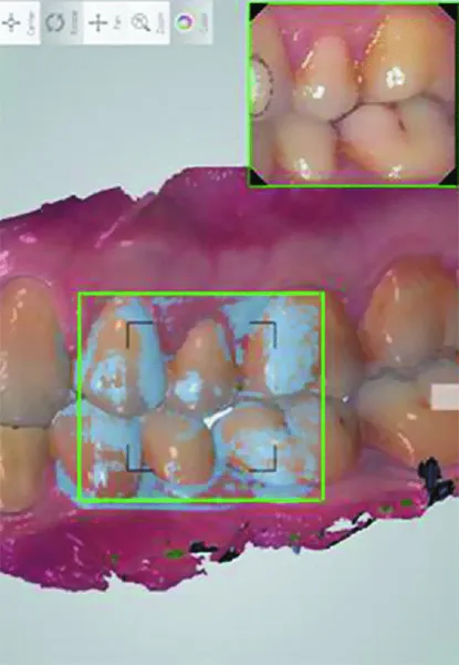
It is easy with the intraoral scanners to scan the interarch relationship. Registration of the occlusion with an intraoral scanner does not require a separate material for bite registration. The occlusion can be quickly, directly, and accurately captured with the intraoral scanner (Figure 1.4). If intraoral scanners are used, the digital dental models are immediately available for diagnosis and analyzing a malocclusion.
Figure 1.4 Scan of the occlusion. Company: 3Shape.
Digital workflow using intraoral scanners
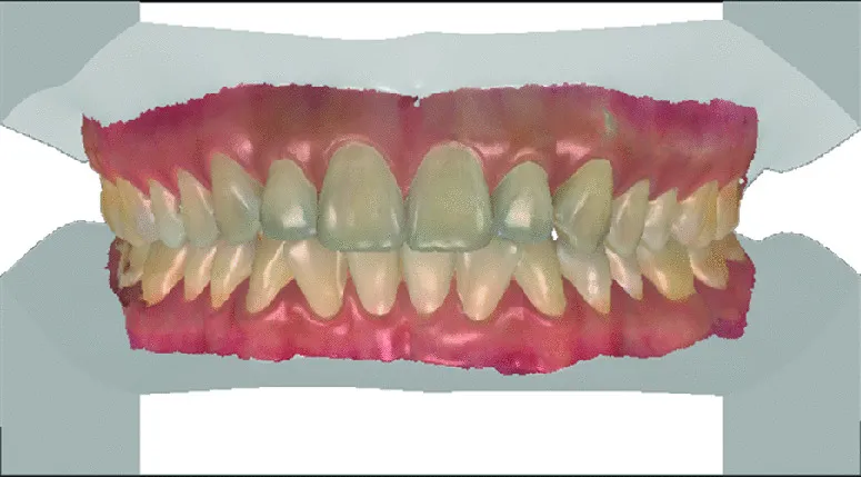
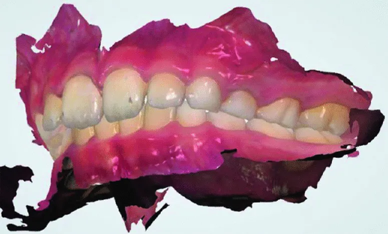
The images of the scanner (some scanners can be used to make a scan, color photographs, and an HD video taken at the same time) have the advantage that they can replace traditional plaster models (Figure 1.5) as well as photography of the dentition (Figure 1.6). Because intraoral scanning is a direct procedure, the intraoral scanning procedure could eventually become more accurate than traditional impression taking as intraoral scanning is not prone to some of the errors that can occur in the traditional impression taking procedure such as air bubbles, rupture of impression material, inaccurate impression tray dimensions, too much or too little impression material, inappropriate adhesion of the impression to the impression tray, and impression material distortion due to the disinfection and transportation procedure.
Figure 1.5 A digital dental model scanned with a color scanner. Company: 3Shape.
Figure 1.6 A intraoral scan in color. Company: 3Shape.
Inaccurate scanning can be improved by rescanning a specific part of the impression, so the procedure to entirely retake an impression can be postponed. Intraoral scanning could be particularly advantageous for patients with anxiety during impression taking (especially for the upper impression) and for cleft palate patients who could carry an increased risk of impression material aspiration and for whom standard impression trays are not suitable. Intraoral scanning could also be an advantage for patients currently undergoing orthodontic treatment with fixed appliances, for whom a traditional impression will be severely distorted because of the presence of the orthodontic appliances. Currently, the mean time needed for intraoral scanning is shorter than that required for taking traditional PVS impressions (one impression with heavy material and a second impression with soft impression material) but longer than required for the alginate impression taking procedure. Most patients have reported that the intraoral scanning procedure is more comfortable than conventional impression taking, especially with PVS impressions, although some studies have reported the opposite conclusion [8–11]. It can be speculated that the reduction in scanning time as well as the possibility to scan without powdering the dentition will improve the positive experience of the patient with the scanning procedure. It is expected that improvem...
Table of contents
Cover
Title Page
Copyright
List of Contributors
Preface
Acknowledgments
1 Documentation of the Dentition
2 Documentation of the Face
3 Dynamic Motion Capture of the Mandible
4 Analysis of Digital Dental Documentation
5 Orthodontic Treatment Planning
6 Custom Appliance Design
7 Custom Appliance Fabrication and Transfer
8 Monitoring of Tooth Movement
9 Custom Retention after Orthodontic Treatment
10 The Invisalign System
11 Custom Lingual Appliances
Appendix
Index
EULA
Frequently asked questions
Yes, you can cancel anytime from the Subscription tab in your account settings on the Perlego website. Your subscription will stay active until the end of your current billing period. Learn how to cancel your subscription
No, books cannot be downloaded as external files, such as PDFs, for use outside of Perlego. However, you can download books within the Perlego app for offline reading on mobile or tablet. Learn how to download books offline
Perlego offers two plans: Essential and Complete
Essential is ideal for learners and professionals who enjoy exploring a wide range of subjects. Access the Essential Library with 800,000+ trusted titles and best-sellers across business, personal growth, and the humanities. Includes unlimited reading time and Standard Read Aloud voice.
Complete: Perfect for advanced learners and researchers needing full, unrestricted access. Unlock 1.5M+ books across hundreds of subjects, including academic and specialized titles. The Complete Plan also includes advanced features like Premium Read Aloud and Research Assistant.
Both plans are available with monthly, semester, or annual billing cycles.
We are an online textbook subscription service, where you can get access to an entire online library for less than the price of a single book per month. With over 1.5 million books across 990+ topics, we’ve got you covered! Learn about our mission
Look out for the read-aloud symbol on your next book to see if you can listen to it. The read-aloud tool reads text aloud for you, highlighting the text as it is being read. You can pause it, speed it up and slow it down. Learn more about Read Aloud
Yes! You can use the Perlego app on both iOS and Android devices to read anytime, anywhere — even offline. Perfect for commutes or when you’re on the go. Please note we cannot support devices running on iOS 13 and Android 7 or earlier. Learn more about using the app
Yes, you can access Digital Planning and Custom Orthodontic Treatment by K. Hero Breuning, Chung H. Kau, K. Hero Breuning,Chung H. Kau in PDF and/or ePUB format, as well as other popular books in Medicine & Orthodontics. We have over 1.5 million books available in our catalogue for you to explore.