In the era of cost cutting and lack of adequate health insurance for many patients, clinical skills and time spent with patients are not adequately compensated. Yet, these dwindling and underpaid skills – good history taking, observation of and listening to patients, and physical examination of patients – remain very essential to making and reaching a complete and accurate diagnosis. Expensive laboratory and imaging diagnostics while very relevant, should not replace these age-old skills that have served to enhance and maintain the doctor-patient relationship and human connection, a connection that is often necessary for healing.
Cases in Clinical Infectious Disease Practice uses case studies to illustrate how the infectious disease clinician processes and integrates data to arrive at a diagnosis. This type of hands-on approach, invaluable in training programs, is utilized to take the reader through initial patient encounter, through the history and physical examination, to simple laboratory findings and stains, to a final diagnosis, in a way that is easily accessible to clinicians, students, and laboratory personnel working with clinical specimens.
Appeals to practitioners of all levels, with focus on patients with common problems or complications of common infections without heavy technical language
Emphasizes basic clinical skills including history taking, observation, epidemiology, and physical exam, as well as simple laboratory tests, explaining how they lead to a reasonable diagnosis
Presents cases seen first-hand within the community setting, reflective of cases or situations a resident or student is likely to encounter in the real world after training
Cases in Clinical Infectious Disease Practice is an essential resource for clinicians, graduate and medical school students, and others conducting medical and clinical microbiology or infectious disease research on real patients.
Trusted by 375,005 students
Access to over 1.5 million titles for a fair monthly price.
Case 1.1 Soft tissue infection following traumatic aquatic exposure
In early September 2010, a 7-year-old boy suffered a laceration injury to the left calf during a recreational boat ride on a coastal river. He was brought to the emergency room (ER) within 2 hours of this accident, after initial cleansing first aid with saline in the field.
The wound was noted to be severe and deep, and was described as “partial degloving” by the ER physician. The boy's vital signs were stable. Due to the nature of the wound, an orthopedic surgeon was consulted and the patient was taken to surgery within 2 hours for wound washout and closure. X-rays of the leg showed no fractures.
Cefazolin and gentamicin were given preoperatively. The patient received 34 stitches to close the large (>10 cm) complex left calf laceration injury after extensive washout. No cultures were done.
One day later, infectious diseases consult was sought for outpatient oral antibiotic recommendations, in anticipation of discharge home later that day.
The boy's past medical history was unremarkable, except for hospital admission for symptoms of nausea and vomiting 1 year earlier, and ear tubes placed 1.5 years earlier.
His examination was unremarkable except for superficial abrasions on the lower abdomen and right upper arm, and the deep (now sutured) left calf laceration. Temperature was 100.5 °F, but other vital signs were stable.
A combination of trimethoprim/sulfamethoxazole and cefuroxime was recommended as oral antibiotics, and the patient was then discharged to outpatient follow-up.
Six days after discharge from the hospital, he was readmitted because of wound infection. He had failed to take the prescribed antibiotics because of severe nausea.
At surgery, no frank pus was found but serous old blood and drainage were noted. A swab of this drainage was stained and cultured.
The infectious diseases consultant was called (after surgery) to help with additional recommendations. He added ceftriaxone to the vancomycin already prescribed. Examination of the patient the next day found him to be comfortable, afebrile, and eating breakfast. His temperature was normal (97.8 °F). The abdominal skin abrasions were healing, but the left calf was wrapped up following the surgical incision and debridement (I&D) the day before.
Basic laboratory findings were normal (white blood cell [WBC] count was 7300/μL, platelet count 401,000/μL, and creatinine 0.5 mg/dL).
The gram stain of the serous fluid from the wound showed rare WBCs and no organisms. Twenty-four hours later, the culture was reported positive for a gram-negative rod (GNR).
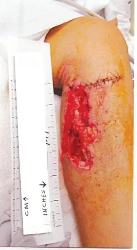
The left calf wound was clean when inspected on day 4 post surgery (Fig. 1.1a).
What are the likely organisms in this patient (differential diagnoses) and why?
Figure 1.1a Left calf wound on day 4 post surgery (reproduced with permission). (See insert for color information)
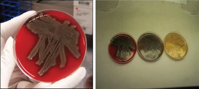
The GNR was found to be oxidase positive, beta-hemolytic on sheep blood agar (BA), and the subculture showed luxuriant growth on all three agar media (BA, chocolate BA, and MacConkey agar) within 24 hours (Fig. 1.1b). It was noted later to be resistant to penicillin/ampicillin-like agents, including carbapenems, but sensitive to second- and third-generation cephalosporins, quinolones, and trimethoprim/sulfamethoxazole, as well as tetracyclines and aminoglycosides.
What is your new diagnosis?
Figure 1.1b Luxuriant growth of gram-negative rod on blood agar medium in 24 hours, and comparative growth of the same organism on BA, chocolate BA, and MacConkey agar. (See insert for color information)
The patient was discharged 6 days later to outpatient follow-up on intravenous ceftriaxone after a peripherally inserted central catheter (PICC line) was placed.
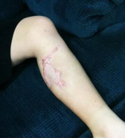
Two months later he was doing very well, and underwent plastic surgical repair of the calf laceration (Fig. 1.1c).
What were the clues to the diagnosis?
Figure 1.1c Photo taken in March 2011, 6 months after the injury: healed left calf post skin grafting (reproduced with permission).
Case discussion
First, the injury occurred while boating on a river, in possibly brackish water (coastal river). Gram-negative organisms are common and likely to contaminate the wound with such a severe laceration injury. The organism grew rapidly on all three culture media used (BA, Choc BA, and MAC). It was oxidase positive, non-lactose fermenting, mucoid, and beta-hemolytic on sheep BA. It did not require high salt concentration to grow, making certain Vibrio organisms (those that need high, 6.5% salt concentration) unlikely. The sensitivity pattern is also useful (see earlier). The organism turned out to be sensitive to the two agents originally recommended (trimethoprim/sulfamethoxazole and cefuroxime). The child was unable to take the antibiotics because of persistent nausea.
Differential diagnosis
Our differential diagnoses at the onset were Aeromonas or Pleisiomonas, more likely than Vibrio, because of the fresh water or brackish water environment where the injury occurred. The luxuriant growth in all three media was in keeping with the differential diagnoses chosen.
Final diagnosis: Aeromonas hydrophila
Some data from the literature on Aeromonas are shown in Table 1.1a and given below.
Table 1.1a Organisms associated with soft tissue infection following water exposure
Organisms
Exposure
Clinical syndromes
Aeromonas spp*
Fresh water
Rapidly developing infection associated with fever; sepsis
Edwardsiella tarda*
Fresh water
Cellulitis, occasionally fulminant infection with bacteremia
Chapter 7: Infectious Diseases Associated with Trauma and Outdoor Activities
Chapter 8: Acute and Chronic Subcutaneous Fungal Infections
Chapter 9: Endocarditis with Unusual Organisms or Characteristics
Chapter 10: Severe Systemic Fungal and Other Infections in AIDS Patients
Chapter 11: Toxic Manifestations of Infectious and Non-infectious Diseases
Chapter 12: Skin and Soft Tissue Infections Seen Post Hurricane Katrina in 2005
Chapter 13: Other Miscellaneous Infections
Index
End User License Agreement
Frequently asked questions
Yes, you can cancel anytime from the Subscription tab in your account settings on the Perlego website. Your subscription will stay active until the end of your current billing period. Learn how to cancel your subscription
No, books cannot be downloaded as external files, such as PDFs, for use outside of Perlego. However, you can download books within the Perlego app for offline reading on mobile or tablet. Learn how to download books offline
Perlego offers two plans: Essential and Complete
Essential is ideal for learners and professionals who enjoy exploring a wide range of subjects. Access the Essential Library with 800,000+ trusted titles and best-sellers across business, personal growth, and the humanities. Includes unlimited reading time and Standard Read Aloud voice.
Complete: Perfect for advanced learners and researchers needing full, unrestricted access. Unlock 1.5M+ books across hundreds of subjects, including academic and specialized titles. The Complete Plan also includes advanced features like Premium Read Aloud and Research Assistant.
Both plans are available with monthly, semester, or annual billing cycles.
We are an online textbook subscription service, where you can get access to an entire online library for less than the price of a single book per month. With over 1.5 million books across 990+ topics, we’ve got you covered! Learn about our mission
Look out for the read-aloud symbol on your next book to see if you can listen to it. The read-aloud tool reads text aloud for you, highlighting the text as it is being read. You can pause it, speed it up and slow it down. Learn more about Read Aloud
Yes! You can use the Perlego app on both iOS and Android devices to read anytime, anywhere — even offline. Perfect for commutes or when you’re on the go. Please note we cannot support devices running on iOS 13 and Android 7 or earlier. Learn more about using the app
Yes, you can access Cases in Clinical Infectious Disease Practice by Okechukwu Ekenna in PDF and/or ePUB format, as well as other popular books in Biological Sciences & Microbiology. We have over 1.5 million books available in our catalogue for you to explore.