X rays are a form of energy belonging to the electromagnetic (EM) spectrum. Some of the members of the EM family include radio waves, microwave radiation, infrared radiation, visible light, ultraviolet radiation,
x‐ray radiation and gamma radiation. These examples are differentiated by their wavelength and frequency. A
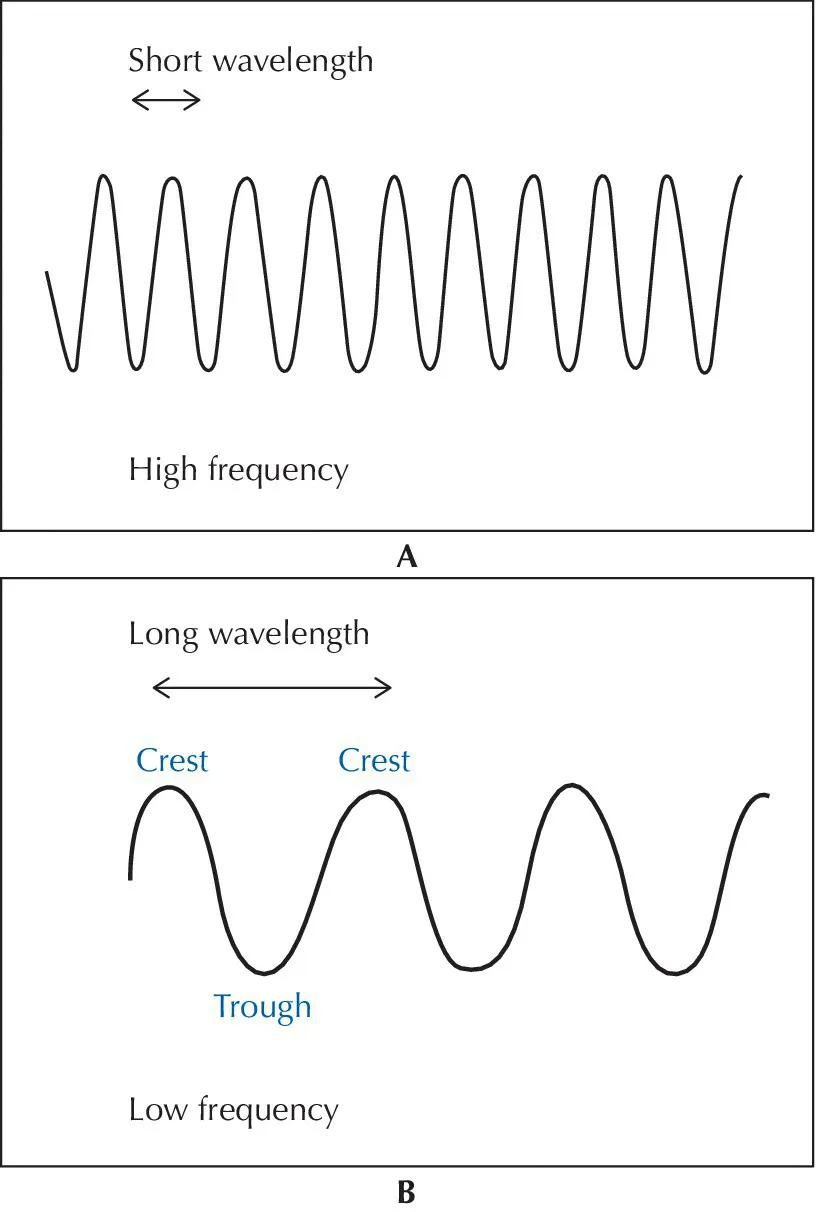
wavelength is defined as the distance between two identical points on consecutive waves (e.g. distance from one crest to the next crest) (
Fig. A1). Longer wavelengths have lower frequencies and are considered to be less damaging to living tissues. Conversely, shorter wavelengths
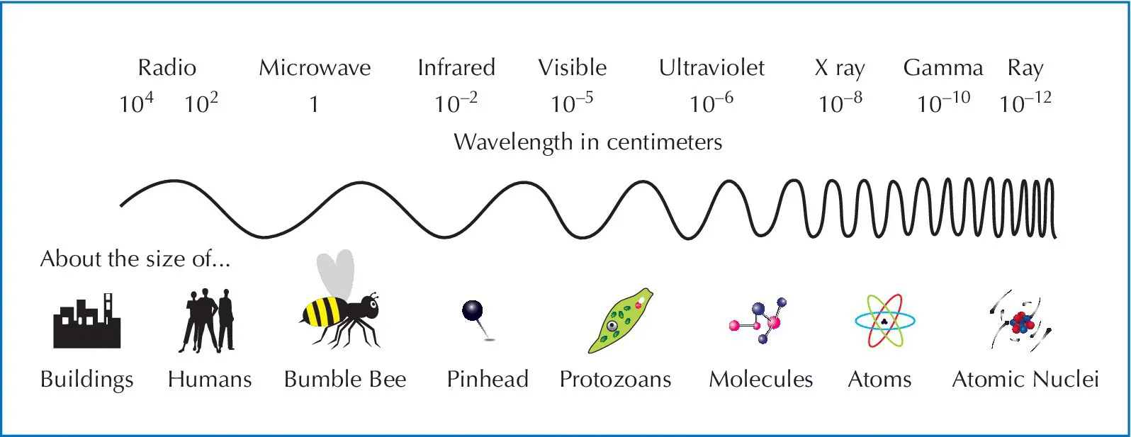
have higher frequencies and are considered to be more damaging to living tissues. One end of the EM spectrum includes the long wavelengths used for radio signal communications while at the short wavelength end of the spectrum is gamma radiation. The EM spectrum covers wavelengths, ranging from
nanometers to kilometers in length (
Fig. A2). Dental x rays are 0.1 to 0.001 nanometers (nm) in length. For comparison purposes, dental x rays may be the size of a single atom while some radio waves are equivalent to the height of a tall building. As with all types of EM radiation, x rays are pure energy. They do not have any mass and because they have very short wavelengths, x rays can easily penetrate and potentially damage living tissues. All forms of EM radiation must not be confused with
particulate radiation, such as
alpha and
beta radiation. Particulate radiation is not discussed in this textbook.
The EM spectrum is divided into the
non‐ionizing forms and the
ionizing forms of radiation. The boundary between non‐ionizing and ionizing radiation is not sharply delineated. Ionizing radiation is considered to begin with the shorter wavelength ultraviolet rays and the increasingly shorter wavelengths which include x rays and gamma rays. The longer wavelengths of ultraviolet rays and beyond which include microwaves, radio waves, etc. are all considered to be non‐ionizing forms of radiation. The difference is that ionizing radiation is powerful enough to knock an
electron out of its atomic orbit, while non‐ionizing radiation is
not powerful enough to remove an electron. The removal of an electron from an atom is referred to as “ionization.” Exposure to ionizing radiation is recognized as being more hazardous to living tissue than non‐ionizing radiation.
In lay terms, x‐ray images reveal the different parts of our bodies or other matter in varying shades of black and white. Why? This is because skin, bone, teeth, fat and air absorb different quantities of radiation. Within the human body, the calcium in bones and teeth absorbs the most x rays. Tooth enamel is the most mineralized substance in the human body (over 90% mineralized). Consequently, mineralized structures such as teeth and bones appear as varying shades of white (i.e.
radiopaque) on dental images. Fat and other soft tissues absorb less radiation, and consequently they will look darker (i.e.
radiolucent) in comparison to bone. Air absorbs the least amount of x rays, so airways and sinuses typically look black in comparison to mineralized substances. The denser or thicker the material, the more x‐ray photons are absorbed by it. This results in a more radiopaque appearance on an x‐ray image. The thinner or less dense an object is, the fewer the number of x‐ray photons absorbed or blocked by it. Thus more x‐ray photons are able to ...