1. Positioning, examining, and opening of the carcass
After the body weight is determined or estimated, the animal is positioned in left lateral recumbency. For identification purposes, a whole-body photograph is taken. If there is a tattoo or brand, these are recorded. The external examination involves all body orifices, mucous membranes including the lining, ocular sclera, the hooves, ears, eyes, and the condition of the hair coat. Valuable information concerning disease of internal organs can be obtained by observing color changes of visible mucous membranes. The gross examination should also include the nutritional status of the animal.
A ventral midline incision extending from the pelvis to the head facilitates the subsequent skinning of the carcass and reflection of the right front leg and hind leg. In the male horse the prepuce is reflected and testes removed from the scrotal sac. This is followed by opening of the abdominal cavity by incision through the abdominal wall.
2. Evisceration of the abdominal, pelvic, and thoracic organs
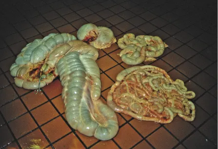
The abdominal viscera, in particular the intestinal tract, are checked for expected anatomic position. The amount and character of the peritoneal fluid (100–200 mL normal, clear, and straw colored) is assessed. After examination of the cranial mesenteric root, the entire gastrointestinal tract is removed to inspect and remove the remaining organs from the abdominal cavity. Individual organs are assessed for color, consistency, symmetry, and, where appropriate, aspect of the cut surface. Individual organs can be weighed and measured under certain circumstances.
The bones of the pelvic cavity are cut with a handsaw for removal of the organs of the pelvic cavity including the urinary bladder, rectum, and genital tract.
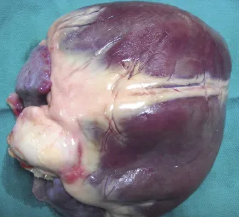
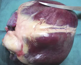
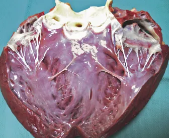
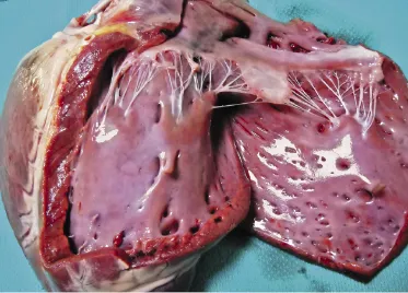
The right rib cage is removed from the chest using strong rib cutters. The entire pluck is removed en bloc, starting with loosening of the tongue from the oral cavity. The tongue is left attached to the esophagus, which is cut at the diaphragmatic hiatus and removed from the opened thorax together with trachea, lungs, and the heart. Trachea, lungs, and heart are separately examined.
a. Cranial mesenteric artery
After dissection of the perivascular tissue, the vascular branch is opened with scissors. The mesenteric arterial root lumen is checked for patency and smoothness of its intima to rule out the presence of Strongylus vulgaris larvae.
3. Removal of the brain and spinal cord
Before removing the head, cerebrospinal fluid can be collected by syringe from the atlanto-occiptal cistern for analysis to aid in the diagnosis of central nervous system disease. Decapitation is performed at the level of the atlanto-occipital joint. The guttural pouches near the occipital condyles are inspected. The cranial cervical ganglion is located next to the carotid artery in the dorsocaudal aspect of the medial compartment of the guttural pouches and can be felt as a bulge in the fold of tissue at that site.
The severed head is placed tightly into a vice or on a solid surface against a rigid object such as a...