Brain–computer interfaces (BCI) are devices which measure brain activity and translate it into messages or commands, thereby opening up many investigation and application possibilities. This book provides keys for understanding and designing these multi-disciplinary interfaces, which require many fields of expertise such as neuroscience, statistics, informatics and psychology.
This first volume, Methods and Perspectives, presents all the basic knowledge underlying the working principles of BCI. It opens with the anatomical and physiological organization of the brain, followed by the brain activity involved in BCI, and following with information extraction, which involves signal processing and machine learning methods. BCI usage is then described, from the angle of human learning and human-machine interfaces.
The basic notions developed in this reference book are intended to be accessible to all readers interested in BCI, whatever their background. More advanced material is also offered, for readers who want to expand their knowledge in disciplinary fields underlying BCI.
This first volume will be followed by a second volume, entitled Technology and Applications.
Trusted by 375,005 students
Access to over 1.5 million titles for a fair monthly price.
This chapter’s objective is not to describe the nervous system in detail, which would be impossible to do in just a few pages, but rather to provide readers who are interested in Brain–Computer Interfaces but who are not an experts in anatomy, with some basics of neuroanatomy and functional anatomy as well as the vocabulary used to talk about them. Readers looking for greater depth and precision in the description of anatomical structures may consult reference books in neuroanatomy (we can cite for their clarity and exhaustiveness [KAM 13, CHE 98, DUU 98])
This description seeks to provide a general understanding of the structure of the adult nervous system, its main constituents and their principal functions, and to thereby better understand the pathologies associated with it.
This chapter will first provide a general description of the nervous system, and it will then focus on a description of the central nervous system (CNS), as well as that of the peripheral nervous system (PNS). In the last section, we will succinctly describe the main pathologies that can be addressed through the use of Brain–Computer Interfaces.
1.1. General description of the nervous system
A neuron is composed of a cell body and an axon, which terminates in a synaptic area. The information that travels through it is an electric signal that corresponds to a depolarization of the axonal membrane: the action potential. In this way, the axon transmits the action potential up to the synapse, the area of communication between neurons. Molecules emitted at the synapses under the influence of action potentials are called neurotransmitters. These neurotransmitters may either be excitatory or inhibitory and thus determine the response obtained.
Neurons are organized in pathways, tracts or networks whose connections determine their roles. Traditionally, a distinction is made between the CNS and the PNS. It is common to talk about efferent neurons, which transmit information from the CNS to the PNS, and afferent neurons, which transmit information from the PNS to the CNS.
The CNS includes the encephalon, which is enclosed in the skull, and the spinal cord in the spinal canal. The encephalon is itself composed of the brain stem, the cerebellum and the two hemispheres of the brain. The brain stem, located in the most caudal part of the encephalon, gives way to 12 pairs of nerves that are known as cranial nerves. The cerebellum is located in the back of the brain stem. Each hemisphere is composed of several lobes (frontal, parietal, temporal, occipital and the insular cortex). From a functional perspective, each hemisphere has its own specific functions, especially for the most complex functions (for example language in the frontal and temporal areas of the dominant hemisphere, spatial orientation in the right parietal lobe, the organization of complex gestures in frontal lobe, etc.).
The cortex, which is located on the surface of the hemispheres, is composed of gray matter that contains neuron cell bodies and is organized into six layers. The basal ganglia are located at the base of the hemispheres. These are also composed of gray matter. White matter contains myelinated axons from CNS neurons and it makes it possible to establish connections between different parts of the CNS through associative fibers (connecting parts of the cortex to each other or to the basal ganglia) and through fibers that stretch out toward the spinal cord.
The spinal cord, which contains ascending fibers and descending fibers, transmits all motor, sensitive and vegetative information between the encephalon and the PNS. It is also composed of gray matter and is the regulation center for a certain number of reflex actions.
The roots that give way to the PNS arise from the spinal cord. These roots form, passing through the (brachial and lumbosacral) plexuses, the entire set of nerve trunks that make it possible to innervate the skeletal muscles (efferent motor fibers) to transmit sensory (sensitive afferent fibers) and vegetative (efferent and afferent vegetative fibers) information.
Different systems (motor, somatosensory, sensory) may have either ascending or descending pathways, going from the peripheral receptor to the area of the brain involved in interpreting the signal, or going from the cortex all the way to the effector (for example the muscle). We may cite, for example, the descending motor tracts distributed in a (corticospinal and corticobulbar) pyramidal pathway, which is the pathway for voluntary motion. We may also cite extrapyramidal pathways, which include other motor pathways. Other pathways include sensitive, visual, auditory, vestibular and olfactory tracts.
1.2. The central nervous system
The CNS includes the encephalon, which is located in the skull, and the spinal cord, which is located in the spinal canal.
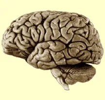
Figure 1.1.General view of the human encephalon (http://lecerveau.mcgill.ca)
The encephalon (Figure 1.1) is usually composed of the following structures:
– the telencephalon;
– the diencephalon;
– the brain stem itself comprising the midbrain, the pons and the medulla oblongata. The cerebellum is located in the back of the pons, which is connected to the pons through the cerebellar peduncle.
It is also possible to describe the encephalon from its formation at the embryonic stage. In such a case, we can distinguish between the hindbrain, which will become the medulla oblongata and the metencephalon (pons and cerebellum), the midbrain and the prosencephalon, which will turn into the diencephalon and the telencephalon.
1.2.1.The telencephalon
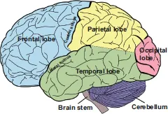
The cerebrum is composed of two hemispheres (right and left) that are connected to one another through white matter tracts (especially by the corpus callosum). The surface of each hemisphere has a folded aspect, which makes it possible to individualize the lobes (Figure 1.2): the frontal lobe, the parietal lobe, the occipital lobe and the temporal lobe on the surface, and the insular lobe on the inside. These lobes are separated by sulci: the central sulcus (also known as the fissure of Rolando), the lateral sulcus or Sylvian fissure, the parietooccipital sulcus and the temporal-occipital sulcus.
Figure 1.2.General view of the cortex’s surface, main lobes and sulci
The surface of each lobe itself includes several convolutions, which are known as gyri, and which make it possible to individualize the most superficial parts of the cortex. Despite variations of this structure among different individuals, it is possible to individualize sulci, fissures and gyri in most subjects with relative constancy either in morphological or functional terms.
Korbinian Brodmann, an early 20th Century neurologist and neuropsychologist, established a map of the cerebral cortex by describing 52 areas based on the tissue and histological composition of the cortex (cytoarchitectonic analysis). These are known as Brodmann areas. Brodmann attributed a specific function to each of them. Some of those areas are now subdivided into subareas, and that mapping is still used today [GAR 06].
The functional role of the different areas of the cerebral cortex is traditionally described in the following manner:
– the primary areas, which include the primary motor cortex, and areas that receive sensory stimuli: primary somatosensory cortex (parietal lobe) for sensory information, primary auditive cortex (temporal lobe) and primary visual lobe (occipital lobe);
– the secondary areas, which correspond to elaborate information processing that may be plurimodal, and associative areas, whose functions are more amodal (cognitive and attentional functions) and that most notably make it possible to pay attention to stimuli to identify them. Cognitive functions are processed in such areas.
Let us now review the different lobes:
–The frontal lobe: The frontal lobe is composed of the precentral gyrus, the premotor areas and the prefrontal areas. In the dominant hemisphere, it contains Broca’s area, which is considered the area of speech production. It is delimited by the central sulcus, which separates it from the parietal lobe, and by the lateral...
Table of contents
Cover
Table of Contents
Title
Copyright
Foreword
Introduction
PART 1: Anatomy and Physiology
PART 2: Signal Processing and Machine Learning
PART 3: Human Learning and Human-Machine Interaction
List of Authors
Index
Contents of Volume 2
End User License Agreement
Frequently asked questions
Yes, you can cancel anytime from the Subscription tab in your account settings on the Perlego website. Your subscription will stay active until the end of your current billing period. Learn how to cancel your subscription
No, books cannot be downloaded as external files, such as PDFs, for use outside of Perlego. However, you can download books within the Perlego app for offline reading on mobile or tablet. Learn how to download books offline
Perlego offers two plans: Essential and Complete
Essential is ideal for learners and professionals who enjoy exploring a wide range of subjects. Access the Essential Library with 800,000+ trusted titles and best-sellers across business, personal growth, and the humanities. Includes unlimited reading time and Standard Read Aloud voice.
Complete: Perfect for advanced learners and researchers needing full, unrestricted access. Unlock 1.5M+ books across hundreds of subjects, including academic and specialized titles. The Complete Plan also includes advanced features like Premium Read Aloud and Research Assistant.
Both plans are available with monthly, semester, or annual billing cycles.
We are an online textbook subscription service, where you can get access to an entire online library for less than the price of a single book per month. With over 1.5 million books across 990+ topics, we’ve got you covered! Learn about our mission
Look out for the read-aloud symbol on your next book to see if you can listen to it. The read-aloud tool reads text aloud for you, highlighting the text as it is being read. You can pause it, speed it up and slow it down. Learn more about Read Aloud
Yes! You can use the Perlego app on both iOS and Android devices to read anytime, anywhere — even offline. Perfect for commutes or when you’re on the go. Please note we cannot support devices running on iOS 13 and Android 7 or earlier. Learn more about using the app
Yes, you can access Brain-Computer Interfaces 1 by Maureen Clerc, Laurent Bougrain, Fabien Lotte, Maureen Clerc,Laurent Bougrain,Fabien Lotte in PDF and/or ePUB format, as well as other popular books in Biological Sciences & Biotechnology. We have over 1.5 million books available in our catalogue for you to explore.