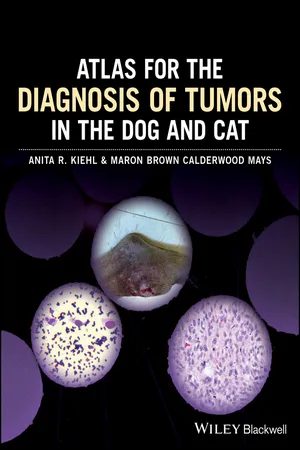
Atlas for the Diagnosis of Tumors in the Dog and Cat is a diagnostic tool for determining if samples are abnormal and defining the cause of the abnormality, with 386 clinical images depicting normal and abnormal results.
Offers a brief overview of the methods used to produce a diagnosis and prognosis from a biopsy tissue sample
Pairs photographs of biopsy samples with photomicrographs of cells obtained via fine needle aspirate
Includes a useful chapter covering sample handling, staining, and shipping
Trusted by 375,005 students
Access to over 1.5 million titles for a fair monthly price.
Tumor is a word of Latin derivation meaning a swelling or protruberance—a “mass.” In its broadest sense it includes masses formed by cellular inflammatory infiltrates, controlled proliferations of hyperplastic cells, and uncontrolled proliferations of neoplastic cells (cancer). Controlled proliferations of cells have a recognizable structure, may perform their usual physiologic function, do not invade local tissues, and suffer senescence and programmed cell death (apoptosis). Uncontrolled proliferations may contain cells of variable structure, may be functional or nonfunctional, may invade local tissues or cause local tissue necrosis by their increasing bulk, replicate in a disorderly manner, and do not suffer programmed cell death.
The path to successful treatment of a tumor begins with recognition of the lesion on a gross level, usually by the caretaker of a dog or cat, sometimes by a groomer, or often during a physical exam by a veterinarian. The next step is assignment of the pathological process into a category of inflammation, hyperplasia, or neoplasia, or some combination of these categories. This can be accomplished at the point of care by aspirating the lesion with a needle and examining the individual cells. In the following chapters this will be called fine needle aspiration (FNA). With some tumors, especially papillomas, impression or scraping of the lesion can yield diagnostic cells, but generally this is not the ideal method of collection, as surface contamination can make evaluation difficult. All cytologic samples are stained with Wright‐Giemsa (W‐G) stain unless otherwise indicated.
Figure 1.1 shows an apocrine gland adenoma FNA. This cluster of small epithelial cells is suggestive of a proliferation of basaloid epithelial cells or the ductular epithelium of an apocrine gland. The cell nuclei are small and regular, and there is scant inflammation, as shown by the neutrophil in this field.
Figure 1.1 Apocrine gland adenoma FNA. 50x.
When FNA of a mass reveals a population of proliferating cells, indicating the lesion is not merely an influx of inflammatory cells that can be relieved by medical means, biopsy allows histopathological evaluation of the affected tissue, showing the architectural arrangement of the cells and allowing for a more definitive diagnosis. This is the point where a hyperplastic growth is distinguished from a neoplastic growth based on how the cells are structurally arranged. FNA cannot evaluate architectural arrangement accurately, because the cells are usually stripped of their association by the process of aspiration. The decision to take an incisional biopsy that removes a portion of the mass, or an excisional biopsy that removes all of the mass, should be based on factors such as the tumor type suggested by the FNA, the size of the lesion, the location of the lesion, the stage of the disease, and other parameters such as the overall health of the patient and wishes of the owner. Ultimately the decision rests on the clinical judgment of the surgeon. All biopsies shown in the following pages are stained with hematoxalin and eosin (H&E), unless otherwise indicated.
Figure 1.2 is a biopsy showing the architecture of the gland aspirated in Figure 1.1. There are double rows of small epithelial cells proliferating in a manner that does not invade into the adjacent stroma, indicating that this is a benign apocrine gland tumor referred to as an apocrine ductular adenoma.
FNA can sometimes identify cells that are so clearly abnormal, either by morphology or cell density, that neoplasia can be diagnosed on a presumptive basis.
Figure 1.3 shows a transitional cell carcinoma FNA. An adult female mixed breed dog was presented for hematuria and dysuria. A tentative diagnosis of cystitis was made based on clinical signs, and cystocentesis was performed to collect urine for routine urinalysis and sedimentation. Cytologic exam revealed many clusters of large epithelioid cells with marked anisokaryosis (variation in nuclear size) and basophilic cytoplasm. There were scattered neutrophils, erythrocytes, and cellular debris. No infectious agents were seen. A preliminary diagnosis of neoplasia, probable transitional cell carcinoma, was made. Treatment for infectious cystitis, based just on clinical signs, would prove useless and would delay the true diagnosis. If neoplasia is suspected on presentation, catheterization would be the preferable method of collect...
Table of contents
Cover
Title Page
Table of Contents
Preface
Acknowledgments
PART I: Overview of the Diagnostic Process
PART II: Case Studies
Index
End User License Agreement
Frequently asked questions
Yes, you can cancel anytime from the Subscription tab in your account settings on the Perlego website. Your subscription will stay active until the end of your current billing period. Learn how to cancel your subscription
No, books cannot be downloaded as external files, such as PDFs, for use outside of Perlego. However, you can download books within the Perlego app for offline reading on mobile or tablet. Learn how to download books offline
Perlego offers two plans: Essential and Complete
Essential is ideal for learners and professionals who enjoy exploring a wide range of subjects. Access the Essential Library with 800,000+ trusted titles and best-sellers across business, personal growth, and the humanities. Includes unlimited reading time and Standard Read Aloud voice.
Complete: Perfect for advanced learners and researchers needing full, unrestricted access. Unlock 1.5M+ books across hundreds of subjects, including academic and specialized titles. The Complete Plan also includes advanced features like Premium Read Aloud and Research Assistant.
Both plans are available with monthly, semester, or annual billing cycles.
We are an online textbook subscription service, where you can get access to an entire online library for less than the price of a single book per month. With over 1.5 million books across 990+ topics, we’ve got you covered! Learn about our mission
Look out for the read-aloud symbol on your next book to see if you can listen to it. The read-aloud tool reads text aloud for you, highlighting the text as it is being read. You can pause it, speed it up and slow it down. Learn more about Read Aloud
Yes! You can use the Perlego app on both iOS and Android devices to read anytime, anywhere — even offline. Perfect for commutes or when you’re on the go. Please note we cannot support devices running on iOS 13 and Android 7 or earlier. Learn more about using the app
Yes, you can access Atlas for the Diagnosis of Tumors in the Dog and Cat by Anita R. Kiehl,Maron Brown Calderwood Mays in PDF and/or ePUB format, as well as other popular books in Medicine & Veterinary Medicine. We have over 1.5 million books available in our catalogue for you to explore.