Atlas of Dermatopathology
  1. English
  2. ePUB (mobile friendly)
  3. Available on iOS & Android
eBook - ePub

About this book

Improve your dermatological diagnostic accuracy.

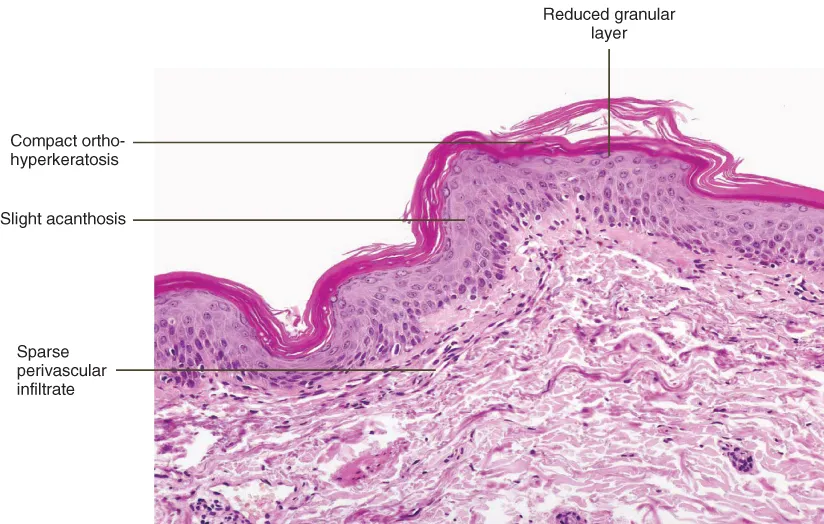
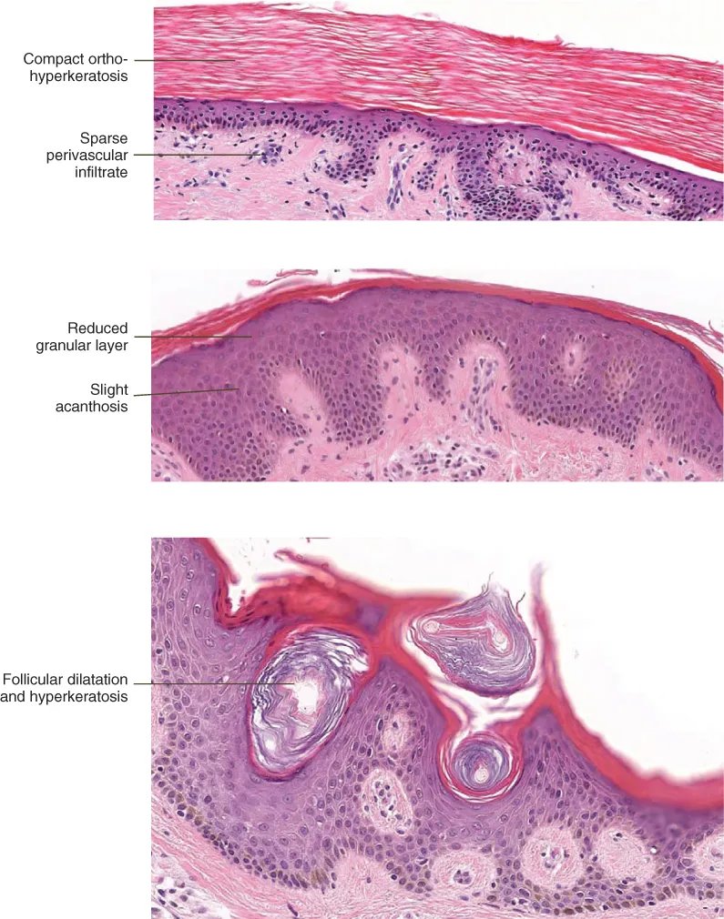
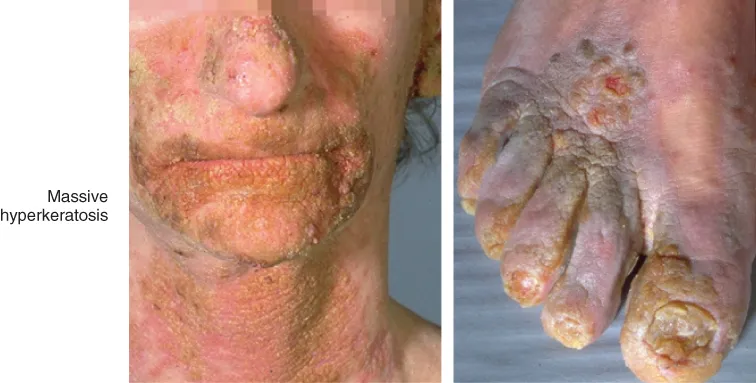
The most efficient method in the diagnosis of skin diseases is the combined validation of clinical and histopathological features. The results of biopsy investigation in isolation don't always yield answers. But when considered together, the clinical pattern and the histopathology, then diagnosis can become clearer. In this process the diagnostic impact of histopathology may be decisive or just confirmative to the clinical differential diagnosis. The structure of the book  follows a basic approach to morphology, which is overall orientation at scanning magnification first, then identifying a prototypic pattern, and finally  finding the essential diagnostic clue(s) under high power magnification.

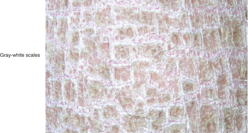
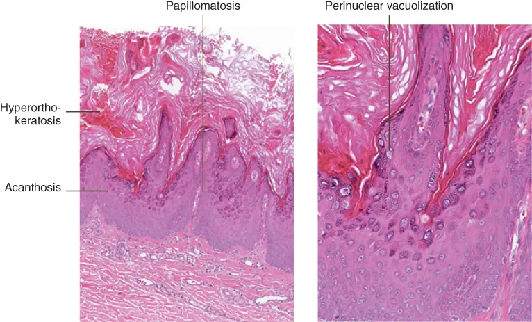
Dermatopathology: Practical Differential Diagnosis by Clinicopathologic Pattern provides top quality images to correlate clinical presentations with histopathologic features. Annotated images highlight subtle indications that can clinch the diagnosis. Concise, bullet-pointed text provides further context.

Written by internationally renowned authors, the book is ideal for anyone involved in the diagnosis of skin disease.  

Frequently asked questions

Yes, you can cancel anytime from the Subscription tab in your account settings on the Perlego website. Your subscription will stay active until the end of your current billing period. Learn how to cancel your subscription.
At the moment all of our mobile-responsive ePub books are available to download via the app. Most of our PDFs are also available to download and we're working on making the final remaining ones downloadable now. Learn more here.
Perlego offers two plans: Essential and Complete
  • Essential is ideal for learners and professionals who enjoy exploring a wide range of subjects. Access the Essential Library with 800,000+ trusted titles and best-sellers across business, personal growth, and the humanities. Includes unlimited reading time and Standard Read Aloud voice.
  • Complete: Perfect for advanced learners and researchers needing full, unrestricted access. Unlock 1.4M+ books across hundreds of subjects, including academic and specialized titles. The Complete Plan also includes advanced features like Premium Read Aloud and Research Assistant.
Both plans are available with monthly, semester, or annual billing cycles.
We are an online textbook subscription service, where you can get access to an entire online library for less than the price of a single book per month. With over 1 million books across 1000+ topics, we’ve got you covered! Learn more here.
Look out for the read-aloud symbol on your next book to see if you can listen to it. The read-aloud tool reads text aloud for you, highlighting the text as it is being read. You can pause it, speed it up and slow it down. Learn more here.
Yes! You can use the Perlego app on both iOS or Android devices to read anytime, anywhere — even offline. Perfect for commutes or when you’re on the go.
Please note we cannot support devices running on iOS 13 and Android 7 or earlier. Learn more about using the app.
Yes, you can access Atlas of Dermatopathology by Günter Burg, Werner Kempf, Heinz Kutzner, Josef Feit, Laszlo Karai, Gunter Burg,Werner Kempf,Heinz Kutzner,Josef Feit,Laszlo Karai,Günter Burg, Gunter Burg, Werner Kempf, Heinz Kutzner, Josef Feit, Laszlo Karai in PDF and/or ePUB format, as well as other popular books in Medicine & Dermatology. We have over one million books available in our catalogue for you to explore.

Information

Year
2015
Print ISBN
9781118658314
eBook ISBN
9781118658291
Edition
1
Subtopic
Dermatology

Chapter 1
Horny Layer

CHAPTER MENU
Reduced granular layer
Prominent granular layer

Reduced granular layer

PROTOTYPE: Ichthyosis vulgaris

image
Cl: Starts in first year of life, dry rough scaly skin, gray-white scales are shed, symmetrical sparing of flexural areas, hyperlinear palms and soles, often atopic dermatitis (50%).
image

Ichthyosis vulgaris

image
Hi: Compact orthohyperkeratosis, granular layer reduced or absent, lack of parakeratosis, follicular dilatation and hyperkeratosis. Epidermis usually normal, sometimes acanthotic or atrophic. No or sparse perivascular infiltrate in the papillary dermis.

VARIANTS: Acquired ichthyosis vulgaris

Histology is identical to ichthyosis vulgaris.

DIFFERENTIAL DIAGNOSIS: Ichthyosis hystrix

image
Cl: Massive, dark, sometimes spiny hyperkeratosis. Various genetic forms exist. Flexures, palms and soles are involved.
image
Hi: Mild hyperorthokeratosis, acanthosis, papillomatosis, elongation of rete ridges. Perinuclear vacuolization of granular and spinous layer keratinocytes, presenting epidermolytic features.

Other Diagnosis

Refsum syndrome (heredopathia atactica polyneuritiformis): Vacuolization of basal and suprabasal keratinocytes (accumulation of phytanic acid; Sudan red stain)X-linked dominant ichthyosis (Harlequin ichthyosis): Clinical features similar to ichthyosis vulgaris, but flexures are involved, undescended testes in 30%. Vacuolization of basal and suprabasal keratinocytes (accumulation of phytanic acid; Sudan red stain)
Lamellar ichthyosis: Genetically heterogeneous disorder, usually present at birth presenting as collodion baby in case of generalized involvement. Erythrodermic and non-erythrodermic forms. Transglutaminase-deficiency in most forms. Histology shows mild to moderate hyperorthokeratosis, stratum granulosum normal or broadened, acanthosis, papillomatosis
Bullous, epidermolytic ichthyosis (bullous form of erythrodermia ichthyosiformis congenitalis): Erythroderma at birth with diffuse blistering and erosions, like burned. Histologically the most striking feature is acanthokeratolysis with epidermal thickening leading to superficial blister formation. Tonofilaments can be seen as dark clumps in a shell-like arrangement around the nucleus
Syndromes of ichthyosis and trichothiodystrophy (Tay syndrome): Additional clinical symptoms and biochemical findings.

References

  1. de Berker, D., W. A. Branford, S. Soucek, and L. Michaels (1993). “Fatal keratitis ichthyosis and deafness syndrome (KIDS). Aural, ocular, and cutaneous histopathology.” Am J Dermatopathol 15(1): 64–9.
  2. de Wolf, K., J. M. Gourdain, G. D. Dobbeleer, and M. Song (1995). “A pa...

Table of contents

  1. Cover
  2. Title Page
  3. Copyright
  4. Table of Contents
  5. Dedication
  6. Preface
  7. Abbreviations
  8. Introduction
  9. Chapter 1: Horny Layer
  10. Chapter 2: Epidermis
  11. Chapter 3: Dermal–epidermal Junction (Interface)
  12. Chapter 4: Dermis
  13. Chapter 5: Vessels
  14. Chapter 6: Subcutis
  15. Chapter 7: Deposition and Storage
  16. Chapter 8: Adnexae
  17. Index
  18. End User License Agreement