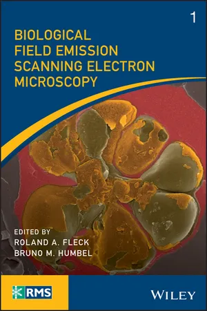
Biological Field Emission Scanning Electron Microscopy
eBook - ePub

Biological Field Emission Scanning Electron Microscopy

Roland A. Fleck, Bruno M. Humbel, Roland A. Fleck, Bruno M. Humbel

  1. English
  2. ePUB (mobile friendly)
  3. Available on iOS & Android
eBook - ePub

Biological Field Emission Scanning Electron Microscopy

Roland A. Fleck, Bruno M. Humbel, Roland A. Fleck, Bruno M. Humbel

Book details
Book preview
Table of contents
Citations

About This Book

The go ? to resource for microscopists on biological applications of field emission gun scanning electron microscopy (FEGSEM)

The evolution of scanning electron microscopy technologies and capability over the past few years has revolutionized the biological imaging capabilities of the microscope—giving it the capability to examine surface structures of cellular membranes to reveal the organization of individual proteins across a membrane bilayer and the arrangement of cell cytoskeleton at a nm scale. Most notable are their improvements for field emission scanning electron microscopy (FEGSEM), which when combined with cryo-preparation techniques, has provided insight into a wide range of biological questions including the functionality of bacteria and viruses. This full-colour, must-have book for microscopists traces the development of the biological field emission scanning electron microscopy (FEGSEM) and highlights its current value in biological research as well as its future worth.

Biological Field Emission Scanning Electron Microscopy highlights the present capability of the technique and informs the wider biological science community of its application in basic biological research. Starting with the theory and history of FEGSEM, the book offers chapters covering: operation (strengths and weakness, sample selection, handling, limitations, and preparation); Commercial developments and principals from the major FEGSEM manufacturers (Thermo Scientific, JEOL, HITACHI, ZEISS, Tescan); technical developments essential to bioFEGSEM; cryobio FEGSEM; cryo-FIB; FEGSEM digital-tomography; array tomography; public health research; mammalian cells and tissues; digital challenges (image collection, storage, and automated data analysis); and more.

  • Examines the creation of the biological field emission gun scanning electron microscopy (FEGSEM) and discusses its benefits to the biological research community and future value
  • Provides insight into the design and development philosophy behind current instrument manufacturers
  • Covers sample handling, applications, and key supporting techniques
  • Focuses on the biological applications of field emission gun scanning electron microscopy (FEGSEM), covering both plant and animal research
  • Presented in full colour

An important part of the Wiley-Royal Microscopical Series, Biological Field Emission Scanning Electron Microscopy is an ideal general resource for experienced academic and industrial users of electron microscopy—specifically, those with a need to understand the application, limitations, and strengths of FEGSEM.

Frequently asked questions

How do I cancel my subscription?
Simply head over to the account section in settings and click on “Cancel Subscription” - it’s as simple as that. After you cancel, your membership will stay active for the remainder of the time you’ve paid for. Learn more here.
Can/how do I download books?
At the moment all of our mobile-responsive ePub books are available to download via the app. Most of our PDFs are also available to download and we're working on making the final remaining ones downloadable now. Learn more here.
What is the difference between the pricing plans?
Both plans give you full access to the library and all of Perlego’s features. The only differences are the price and subscription period: With the annual plan you’ll save around 30% compared to 12 months on the monthly plan.
What is Perlego?
We are an online textbook subscription service, where you can get access to an entire online library for less than the price of a single book per month. With over 1 million books across 1000+ topics, we’ve got you covered! Learn more here.
Do you support text-to-speech?
Look out for the read-aloud symbol on your next book to see if you can listen to it. The read-aloud tool reads text aloud for you, highlighting the text as it is being read. You can pause it, speed it up and slow it down. Learn more here.
Is Biological Field Emission Scanning Electron Microscopy an online PDF/ePUB?
Yes, you can access Biological Field Emission Scanning Electron Microscopy by Roland A. Fleck, Bruno M. Humbel, Roland A. Fleck, Bruno M. Humbel in PDF and/or ePUB format, as well as other popular books in Physical Sciences & Science History & Practice. We have over one million books available in our catalogue for you to explore.

Information

Publisher
Wiley
Year
2019
ISBN
9781118663264

1
Scanning Electron Microscopy: Theory, History and Development of the Field Emission Scanning Electron Microscope

David C. Joy
232 Science and Engineering Research Facility, Department of Materials Science and Engineering, University of Tennessee, Knoxville, TN, USA

1.1 The Scanning Electron Microscope

Since its initial development (Everhart and Thornley, 1958) the scanning electron microscope (SEM) has earned a reputation for being the most widely used, high performance, imaging technology that is available for applications ranging from imaging, fabrication, patterning, and chemical analysis, and for materials of all types and applications. It is estimated that 150 000 or so such instruments are now currently in use worldwide, varying in performance and complexity from simple desk‐top systems to state‐of‐the‐art field emission gun systems that can now cost in excess of $5 million.
The basic principle of the scanning electron microscope is simple. An incident electron beam is brought to a focus that typically varies in size from a fraction of a centimeter in diameter down to a spot that can be smaller by a factor of many thousands of times, and with an energy varying from 100 eV or less to a maximum of 30 keV or more. This beam spot is typically then scanned (Figure 1.1) in a linear “raster” pattern across the region of interest, although other patterns – such as a radial beam – are sometimes employed for special purposes. Typically the final deposited pattern will contain of the order of 1000 × 1000 or more individual imaging points.
Diagram of the SEM scan raster with parts labeled. Arrows link Illumination to Specimen, to Detector outside the SEM, then to Image in the Monitor. On the left list functions of electron gun, condenser 2, and objective.
Figure 1.1 The SEM scan raster.
The incident beam electrons can interact with the sample atoms through either elastic or inelastic scattering. Elastic scattering is where the incident electrons are deflected with no loss of energy. Inelastic scattering involves a loss of energy, often by ionizing the sample atoms. The incident electrons will scatter (both elastically and inelastically) many times in a region of the sample known as the interaction volume. The size of the interaction volume will depend on the incident energy and the nature of the sample, but can be of the order of a micrometer in diameter. A number of different types of signal generated by the beam–sample interaction can be detected. The intensity of the signal detected can be plotted as a function of probe position to form an image. Two important signals are secondary electrons (SEs) and back‐scattered electrons (BSEs). Secondary electrons are electrons from the sample atoms that are released through ionization. They are relatively low in energy <∌25 eV and tend to only escape from the top few tens of nanometers of the surface. They provide strong topographical imaging of surfaces. Back‐scattered electrons are incident electrons that have been multiply scattered and emerge again from the surface. The strength of the scattering that can return the electrons to the surface depends strongly on atomic number, Z, and so BSE imaging gives compositional contrast. Another common signal detected is X‐rays from the decay of the ionized atoms. The energy of the X‐ray photon emitted is characteristic of the element ionized, and so energy‐dispersive X‐ray (EDX) spectroscopy allows mapping of element species.
Most modern SEMs will likely have, and make use of, several types of detector so as to optimally detect, capture, collect, and display other analytical and imaging modes as desired.
In operation the electron source must be carefully set up and optimized so as to generate the smallest spot size for the electrons while still ensuring that the beam current reaching the specimen is adequately stable for periods of many hours without the need for any further operator interactions. The overall measure of imaging performance for the electron source is determined by its brightness ÎČ, which is defined as
equation
where d is the diameter of the spot size of the beam at the target, I is the incident beam current, and α is the solid angle subtended by the illumination at the specimen.
For an electron beam source of some specified energy the beam brightness is said to be “conserved”, which means that varying the beam current – as, for example, by varying the beam spot size or the convergence angle of the beam – will always result in compensating changes in the other parameters in the system so that the magnitude of ÎČ in the equation remains constant. As a result, the intensity of the incident beam current I varies as d 2 α 2 and if either the beam spot size d or the beam convergence angle α are reduced, then the beam current will decrease, which may ultimately result in the beam becoming lost in the background noise of the instrument. The imaging performance of an SEM is very important and therefore is always very dependent on optimum alignment.

1.2 The Thermionic GUN

For the first 25 years or so of the SEM era the only available sources of the energetic electrons required for microscopy were the so‐called thermionic (“hot beam”) emitters mentioned above. Even today so‐called “table‐top” SEM instruments remain in widespread use because of their low cost, good resolution, and operating convenience.
In operation the required electron beam current is generated by heating a tungsten wire filament. This so‐called “thermionic emitter” is usually fabricated from high quality tungsten wire that has been bent into a “V” shape and is maintained at a temperature of about 2700 K by means of a separate power supply that heats the tip region. The “V” shape noted above is maintained at some negative voltage typically from about ∌1 keV – 30 keV with reference to ground potential. The corresponding incident beam currents typically can vary from 10−6 down to 10−12 amps or so.
To optimize the yield of the emitted beam current that is generated a “grid cap”, or a “Wehnelt” cylinder – with a circular aperture centered on the tip of the emitted beam current– is employed. The cap is maintained in position by a potential source that is set to about 50 volts or greater, so that the emitted beam from the source can be brought to a focused crossover at a point chosen some distance beyond the column grid cap. The generated electron beam can then be accelerated down the column and on to the specimen...

Table of contents

Citation styles for Biological Field Emission Scanning Electron Microscopy

APA 6 Citation

[author missing]. (2019). Biological Field Emission Scanning Electron Microscopy (1st ed.). Wiley. Retrieved from https://www.perlego.com/book/993765/biological-field-emission-scanning-electron-microscopy-pdf (Original work published 2019)

Chicago Citation

[author missing]. (2019) 2019. Biological Field Emission Scanning Electron Microscopy. 1st ed. Wiley. https://www.perlego.com/book/993765/biological-field-emission-scanning-electron-microscopy-pdf.

Harvard Citation

[author missing] (2019) Biological Field Emission Scanning Electron Microscopy. 1st edn. Wiley. Available at: https://www.perlego.com/book/993765/biological-field-emission-scanning-electron-microscopy-pdf (Accessed: 14 October 2022).

MLA 7 Citation

[author missing]. Biological Field Emission Scanning Electron Microscopy. 1st ed. Wiley, 2019. Web. 14 Oct. 2022.