An unprecedented book that discusses a decades long journey of understanding vision and visual impairment through working with patients with brain damage
Edward de Haan, a noted clinical vision researcher for the last 35 years, explains how the healthy brain deals with visual information and reveals how he learned to appreciate what it means to be visually impaired. Through discussions of fascinating case studies, he shows that visual deficits are individually unique. Some patients perceive the world without color, some see objects in a distorted manner, whilst others will claim that they can still see although they are demonstrably blind.
The author details his experiences with these patients to demonstrate the manner in which patient work is a unique and vital part of discovering how the brain processes visual information. In doing so, Impaired Vision offers a review of the clinical symptoms related to visual impairment and highlights that the patient study method has not lost any of its relevance in our increasingly high-tech world. This important book:
Explores the various clinical phenomena in visual impairment after brain damage
Demonstrates the effectiveness of the patient study method for understanding visual deficits after brain damage
Contains comprehensive coverage of the variety of symptoms that are manifest in patients with visual impairment
Includes compelling case studies of visually impaired patients
Written for a general audience but of interest for students, researchers and clinicians, Impaired Vision contains fascinating case studies that offer an understanding of the symptoms that are associated with visuals deficits of brain damage.
Trusted by 375,005 students
Access to over 1.5 million titles for a fair monthly price.
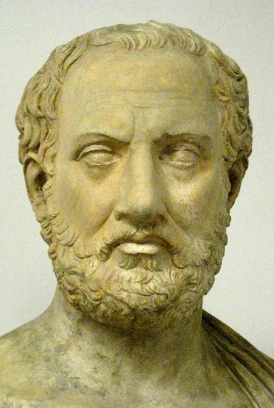
Initially, the structure of the brain was studied in crude ways by dissecting the corpses of animals and humans. Over centuries, more detailed means of investigation were developed, helped by technological advances such as the microscope. The first observations that gave insights into the functional organization came from literary descriptions of people who had suffered brain damage. The following quote comes from the Greek general Thucydides (Figure 1.1), who reported on the effects of the bubonic plague in roughly 400 BC:
Figure 1.1 Bust of the Greek general Thuycidides.
Source: user:shakko. https://commons.wikimedia.org/w/index.php?curid=5573987. Licensed under CC BY‐SA 3.0.
The disease first settled in the head, went on to affect every part of the body in turn, and even when people escaped its worst effects, it still left its traces on them by fastening upon the extremities of the body. It affected the genitals, the fingers, and the toes, and many of those who recovered lost the use of these members; some, too, went blind. There were some also who, when they first began to get better, suffered from a total loss of memory, not knowing who they were themselves and being unable to recognize their friends.
(Thucydides, The History of the Peloponnesian War, Book II, 49, 153)
From this short description, it appears that memory functions may become disrupted after disease. It is also clear that the memory problems may be selective. Some of the survivors were able to talk to Thucydides, so in these patients, speech must have been preserved to a degree. It is conjecture, of course, but it appears that Thucydides was confronted with a large number of patients suffering from a severe brain disease, and he observed that language and memory are separate mental abilities. He had stumbled on an important principle: some functions may become disrupted selectively while others remain intact. He could not guess where the responsible damage in the body – or in the brain, for that matter – was located. He had no means to assess the internal physical locus of the problem.
The opposite was true for the early anatomists, like Galen (second century) and Vesalius (sixteenth century), who were able to look inside the head (post mortem) and who created more and more precise drawings of the brain. Despite their detailed observations, they were somewhat at a loss about functional organization. They could see where the nerve fibers from the sensory systems (e.g., eyes and ears) reached the brain, and inferred correctly that those parts were involved in perception. They were also correct in concluding that motor planning took place in the areas where the nerve fibers that innervate the muscles originate. Apart from these “input” and “output” systems, however, it was extremely difficult for them to determine what the different parts of the brain were doing. For a long time, it was almost impossible to study brain structure and brain function simultaneously. In order to understand how the brain really works, these two approaches needed to be brought together.
One of the first to try to do this was the much vilified Franz Joseph Gall (Figure 1.2). He started his quest for function localization, which he called phrenology, in the late eighteenth century, without a means of looking inside the head. Instead, he suggested that we could use the shape of the head to infer the degree to which a person possessed a certain mental ability. His reasoning was as follows. First, he postulated that all our mental abilities are carried out in the brain (and not, for instance, also in the heart, as many argued at the time). Second, he suggested that specific abilities, such as memory and speech, are carried out in dedicated parts of the brain, which he called “organs.” In addition, he suggested that the size of each organ depends directly on the degree to which a person possesses its associated ability. Thus, by using the analogy of the muscle – more muscle mass means more power – he argued that someone with the “gift of the gab” will have a large language organ in his or her brain. As this particular organ is positioned at the front of the brain, he further suggested that such a person will have protruding eyes. This last suggestion, that the shape of the head gives away detailed information regarding mental abilities and personality traits, such as trustworthiness or patriotic tendencies, was actually not proposed by Gall at all. It is called physiognomics and was popularized by Johann Kaspar Lavater in the second half of the eighteenth century, although its origins can be traced back to Aristotle. Physiognomics has not stood the test of time, not in the least because of the more and more outrageous claims made by its practitioners. For instance, a low forehead, high cheekbones, and a flat nose were all supposed to be signs of aggression, although it was clear that these features often occur in people who are, not aggressive at all. In addition, the descriptions became more and more frivolous, such as facial signs for agility and cynicism.
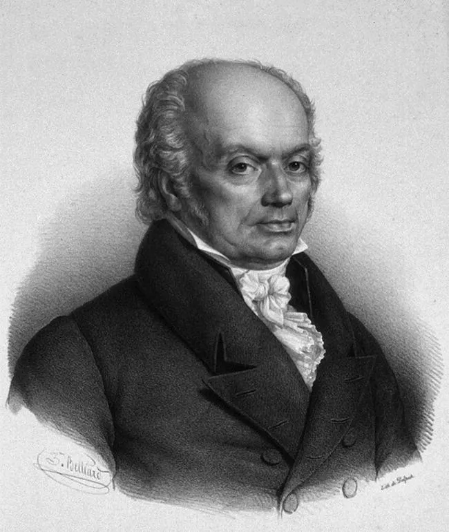
Gall's phrenology was rejected ultimately not because of his use of physiognomics but by early physiological experiments. Gall's proposals were reviewed for the Académie Française by a committee chaired by the pioneer physiologist, Marie Jean Pierre Flourens (1794–1867). Flourens had carried out experiments in which he made surgical lesions in the brains of living rabbits and pigeons, and carefully observed the effects on their behavior. He concluded that, irrespective of the location of the lesion, all functions were affected to an equal degree and that the size of the lesion determined the severity of the impairments. His observations led to the doctrine of “Action Propre”: the position that the brain functions as a whole and that there is no differentiation of function. Gall's scientific career was finished.
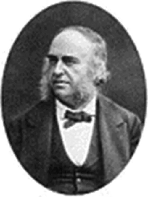
It was not until the second half of the nineteenth century that the combined structural and functional investigation of the human brain commenced in earnest. Neurology developed rapidly as a new discipline within the medical arena in many academic centers in Europe, most notably Paris. Paul Broca (1794–1867, Figure 1.3) was a leading researcher who advocated the careful observation of brain‐damaged patients, followed by detailed post‐mortem examination of their brains. This research method demands great diligence, determination, and creativity. Psychology was in its infancy, and the description of mental abilities was still uninformed. Therefore, these clinicians had to discover new ways of describing the impaired behavior of their patients. Subsequently, they had to wait sometimes years until the patient died in order to carry out an autopsy. Finally, they had to find a conceptual framework for relating impaired functions to the damage they observed in the brain. They were neurologists, psychologists, and anatomists in one, initially without a clear theory of how the brain works.
These scientists learned fast. Broca described a patient, Tan, who was unable to speak as a result of damage to the front of the left part of his brain, or left hemisphere. Later, Carl Wernicke (1848–1905) reported on a patient with damage to the middle of the left hemisphere who was poor at understanding spoken language. Wernicke compared his patient with Broca's, and subsequently did two important things. First, he concluded that these two patients, with different language problems and different lesions, showed that in healthy people language perception and language production are separate processes located in different parts of the brain. Second, he made a prediction. He argued that these two language areas in the brain must be connected, and that if the connection were to be selectively cut by focal brain damage, the patient would show no language perception or speech problems but would be unable to repeat accurately what was said to him or her (see Figure 1.4). That was a bold prediction, but, lo and behold, in 1885, Ludwig Lichtheim (1845–1928) reported exactly such a patient. This was the beginning of a long tradition – a tradition that was to build the first theories of function localization, and a tradition that has lost none its relevance.
Figure 1.4 First schema of language functions in the brain, by Carl Wernicke.
Source: M. Catani & M. Mesulamb (2008). The arcuate fasciculus and the disconnection theme in language and aphasia: History and current state. Cortex, 44(8), 953–961. Reproduced with permission of Elsevier.
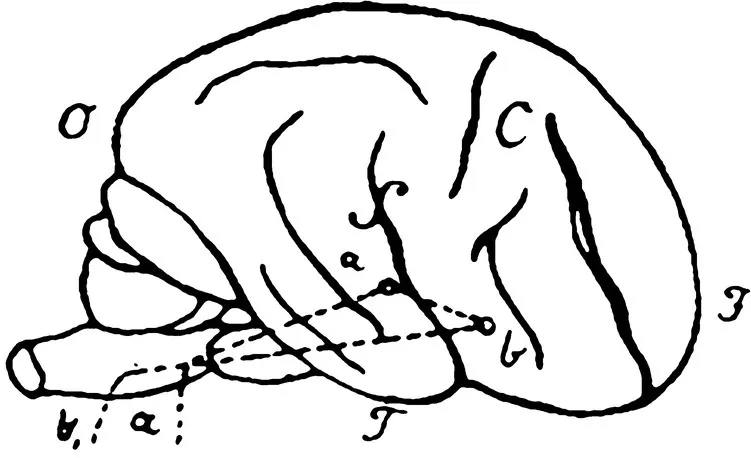
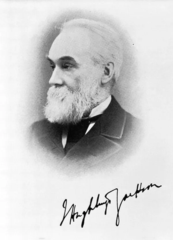
But now back to vision. The early nineteenth‐century explorations of brain function revealed a number of patients with surprising visual impairments. For instance, the English neurologist, John Hughlings Jackson (1835–1911, Figure 1.5), was the first to describe a patient with what he called “imperception.” Following damage to the posterior part of both sides of the brain, this patient was unable to recognize objects, faces, or text, although she was obviously still able to see: she could point to targets and could easily move around in a furnished room without collisions. Hughlings Jackson suggested that, although the visual signals from her eyes did reach her brain and she could still see in a way, the patient was no longer able to recognize the world around her. Everything was new to her. This was the first in a long series of patients with visual problems, stretching into the present. These patients, and the scientists who investigated their visual impairments, beginning with Hughlings Jackson, have provided us with the basic understanding of our visual brain.
Figure 1.5 John Hughlings Jackson.
Source: Wellcome Collection. https://wellcomecollection.org/works/qv6rwm3p?query=L0000492&wellcomeImagesUrl=GET%20/indexplus/image/L0000492.html%20HTTP/1.1. Licensed under CC BY 4.0.
In 1878, Hughlings Jackson started the pres...
Table of contents
Cover
Table of Contents
Preface
1 Looking at the Brain
2 Blind
3 Partially Blind
4 Looking but Not Seeing
5 Seeing Things Differently
6 Seeing What Is Not There
7 Knowing the Unseen
8 Oblivion
9 Vision
Index
End User License Agreement
Frequently asked questions
Yes, you can cancel anytime from the Subscription tab in your account settings on the Perlego website. Your subscription will stay active until the end of your current billing period. Learn how to cancel your subscription
No, books cannot be downloaded as external files, such as PDFs, for use outside of Perlego. However, you can download books within the Perlego app for offline reading on mobile or tablet. Learn how to download books offline
Perlego offers two plans: Essential and Complete
Essential is ideal for learners and professionals who enjoy exploring a wide range of subjects. Access the Essential Library with 800,000+ trusted titles and best-sellers across business, personal growth, and the humanities. Includes unlimited reading time and Standard Read Aloud voice.
Complete: Perfect for advanced learners and researchers needing full, unrestricted access. Unlock 1.5M+ books across hundreds of subjects, including academic and specialized titles. The Complete Plan also includes advanced features like Premium Read Aloud and Research Assistant.
Both plans are available with monthly, semester, or annual billing cycles.
We are an online textbook subscription service, where you can get access to an entire online library for less than the price of a single book per month. With over 1.5 million books across 990+ topics, we’ve got you covered! Learn about our mission
Look out for the read-aloud symbol on your next book to see if you can listen to it. The read-aloud tool reads text aloud for you, highlighting the text as it is being read. You can pause it, speed it up and slow it down. Learn more about Read Aloud
Yes! You can use the Perlego app on both iOS and Android devices to read anytime, anywhere — even offline. Perfect for commutes or when you’re on the go. Please note we cannot support devices running on iOS 13 and Android 7 or earlier. Learn more about using the app
Yes, you can access Impaired Vision by Edward de Haan in PDF and/or ePUB format, as well as other popular books in Biological Sciences & Neuroscience. We have over 1.5 million books available in our catalogue for you to explore.