
- English
- ePUB (mobile friendly)
- Available on iOS & Android
eBook - ePub
Histologic Basis of Ocular Disease in Animals
About this book
Histologic Basis of Ocular Disease in Animals is a comprehensive reference covering pathology of the eye in a spectrum of animal species, including domestic animals, fish, birds, and laboratory animals.
- Offers a comprehensive resource on diseases and conditions of the eye and orbit in a wide range of species
- Covers domestic animals, fish, birds, and laboratory animals
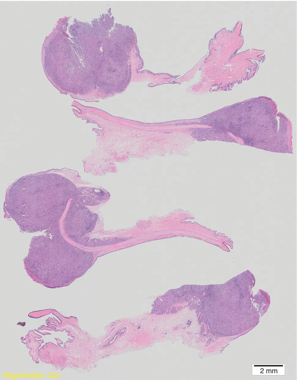
- Presents more than 1200 high-quality images carefully selected to illustrate the ocular conditions covered
- Emphasizes unique pathological responses where necessary
Frequently asked questions
Yes, you can cancel anytime from the Subscription tab in your account settings on the Perlego website. Your subscription will stay active until the end of your current billing period. Learn how to cancel your subscription.
At the moment all of our mobile-responsive ePub books are available to download via the app. Most of our PDFs are also available to download and we're working on making the final remaining ones downloadable now. Learn more here.
Perlego offers two plans: Essential and Complete
- Essential is ideal for learners and professionals who enjoy exploring a wide range of subjects. Access the Essential Library with 800,000+ trusted titles and best-sellers across business, personal growth, and the humanities. Includes unlimited reading time and Standard Read Aloud voice.
- Complete: Perfect for advanced learners and researchers needing full, unrestricted access. Unlock 1.4M+ books across hundreds of subjects, including academic and specialized titles. The Complete Plan also includes advanced features like Premium Read Aloud and Research Assistant.
We are an online textbook subscription service, where you can get access to an entire online library for less than the price of a single book per month. With over 1 million books across 1000+ topics, weâve got you covered! Learn more here.
Look out for the read-aloud symbol on your next book to see if you can listen to it. The read-aloud tool reads text aloud for you, highlighting the text as it is being read. You can pause it, speed it up and slow it down. Learn more here.
Yes! You can use the Perlego app on both iOS or Android devices to read anytime, anywhere â even offline. Perfect for commutes or when youâre on the go.
Please note we cannot support devices running on iOS 13 and Android 7 or earlier. Learn more about using the app.
Yes, you can access Histologic Basis of Ocular Disease in Animals by Bruce Grahn,Robert Peiffer,Brian Wilcock in PDF and/or ePUB format, as well as other popular books in Medicine & Veterinary Medicine. We have over one million books available in our catalogue for you to explore.
Information
1
Fixation and processing of ocular tissues
Fixed ocular tissues are submitted as (i) whole globes with attached orbital tissues from exenteration or transpalpebral enucleation (Figures 1.0 and 1.1); (ii) globes without orbital tissues (transconjunctival enucleation) (Figure 1.2); (iii) intraocular contents from evisceration specimens (Figures 1.3 and 1.4); (iv) prolapsed tissues following penetrating trauma; or (v) excisional or incisional biopsies of proliferative lesions (Figure 1.5). The clinical pathologist receives exfoliative (scrapings or impression smears) or aspirated specimens. Each of these samples has unique preparation requirements in order to optimize the pathologistâs examination (Torcyznski, 1981).

Figure 1.0 A bisected canine globe that was exenterated due to a large retrobulbar tumor that is indenting the sclera (black arrows), the retina is detached (gray arrows) and the vitreous is compressed and contains asteroid hyaloid bodies (white arrow) (formalin fixation).

Figure 1.1 This bisected canine globe has endophthalmitis and was removed by transpalpebral enucleation. This globe contains white purulent exudate, and the iris is extending into a corneal defect (white arrows). Unless they are likely to contain lesions, the eyelids and other periocular soft tissues would ordinarily be removed during transconjunctival enucleations prior to fixation or if they are left on (transpalpebral enucleation and exenterations), they may be trimmed when the globe is sectioned (formalin fixation).

Figure 1.2 A bisected canine globe removed by transconjunctival enucleation, the technique used for the vast majority of globes submitted for histologic assessment. The retina is detached (white arrows) and noticeably thin (formalin fixation).

Figure 1.3 A canine evisceration sample bisected in preparation for histologic processing. Many are not as cohesive as this and arrive as multiple disconnected fragments. Evisceration samples like this should be sectioned in multiple planes to ensure that all intraocular tissues are present for histologic examination (retina, uvea, and lens) (formalin fixation).

Figure 1.4 Lowâmagnification histologic image of a typical evisceration specimen. When contrasted to sections from enucleated globes, such samples provide limited (albeit sometimes still useful) diagnostic information. The predominance of pigmented uveal tissue of which the ciliary body is most abundant is routine (hematoxylin and eosin stain, formalin fixation, and subgross magnification).

Figure 1.5 Lowâmagnification histologic section of a formalinâfixed third eyelid tumor in a dog, confirmed histologically as a minimally invasive adenocarcinoma arising from the gland of the third eyelid (hematoxylin and eosin stain, formalin fixation, subgross magnification).
Tissues other than globes are generally fixed as is routine for nonocular tissues with 10% neutral buffered formalin, while cytology specimens should be submitted as airâdried smears on glass slides. Globes require greater attention; it is important that they be fixed promptly to ensure rapid fixation of the internal structures and minimize autolytic changes. The ideal fixative would (i) penetrate cornea and sclera rapidly; (ii) preserve normal tissue relationships; (iii) allow for detailed gross examination; (iv) minimize embedding, trimming, and cutting artifacts; (v) enable any number of histochemical or immunohistochemical stains; and (vi) permit ultrastructural evaluation in those admittedly few cases in which it might be necessary. As no such perfect fixative exists, selection becomes a compromise influenced by availability, ease of use, and the quality of fixation required for the specific application (Yanoff et al., 1965; Yanoff and Fine, 1967; Yanoff, 1973; Menocal et al., 1980; Margo and Lee, 1995).
Fixatives
The most common method of fixation for light microscopic evaluation of globes is immersio...
Table of contents
- Cover
- Table of Contents
- Foreword
- 1 Fixation and processing of ocular tissues
- 2 General pathology of the eye
- 3 Congenital anomalies
- 4 Histopathology of ocular trauma
- 5 Diseases of the eyelid, conjunctiva, lacrimal, and nasolacrimal systems
- 6 Diseases of the cornea
- 7 Diseases of the episclera and sclera
- 8 Histologic manifestations of disorders of the uvea
- 9 Histologic basis of glaucoma
- 10 Histologic manifestations of acquired and inherited diseases of the lens
- 11 Acquired diseases of the vitreous
- 12 Histologic manifestations of retinal disease
- 13 Acquired diseases of the optic nerve
- 14 Acquired diseases of the orbit
- 15 Intraocular neoplasia
- Index
- End User License Agreement