- Tom Kenny, one of the best-known and well-respected educators in EP brings his signature style to this new primer
- Practical, accessible, highly illustrated approach makes learning easy
- Provides an overview of the algorithms and devices offered by the world's five pacemaker manufacturers
- Offers clinicians learning objectives, test questions and essential points in bulleted lists
- Perfect introductory guide to the topic, assumes little baseline knowledge and appropriate for residents, fellows, EP nurses, general clinical cardiologists, EP fellows and industry professionals

- English
- ePUB (mobile friendly)
- Available on iOS & Android
eBook - ePub
About this book
Trusted by 375,005 students
Access to over 1.5 million titles for a fair monthly price.
Study more efficiently using our study tools.
Information
Chapter 1
Cardiovascular anatomy and physiology
Learning objectives
- Point out the key landmarks in the human heart relevant to cardiac rhythm management.
- Name the four chambers of the heart, the four valves, and the major vessels.
- Describe the flow of blood through the heart.
- Define AV synchrony and explain why it is important.
- State the difference between the body’s arterial versus venous systems.
Introduction
An encyclopedia could be written on the anatomy and physiology of the human heart, and that is not our purpose. Device clinicians must understand the cardiovascular system to understand arrhythmias and device therapy. This chapter will introduce the important concepts of cardiac anatomy and physiology necessary for an understanding of cardiac rhythm management. To that end, this chapter will describe the chambers, valves, and major vessels of the heart and how these control the flow of blood in the body. Although we think of the heart—rightly—as a pump, it also possesses a complex electrical system. The cells of the human heart are unique in many ways, and how they produce, conduct, and dissipate electrical energy is very important, particularly to pacing. Our goal here is to describe the anatomy and physiology of the healthy heart and cardiovascular system in terms of what device clinicians need to know.
The healthy heart
The human heart is a double pump (right and left) that sits in the middle of the chest, slightly to the left, and rotated so that the right side is more anterior than the left. An average adult human heart is relatively large, about 13 by 9 by 6 cm and weighing about 300 g. The heart is protected by the rib cage and sits directly behind one of the body’s thickest bones, the sternum. The bottom of the heart rests on the diaphragm muscle. The heart is encased in this protected but somewhat crowded area—it also contains the lungs (three lobes on the right, two on the left), the stomach, and the intestines.
The bottom tip of the heart (called the apex) taps up against the chest when the heart contracts. By placing his hands on the chest, a physician can feel the place where the apex of the heart makes contact with the chest; this place is called the point of maximal impulse (PMI). Knowing the precise location of the PMI can be very useful in treating cardiology patients, because the PMI of a healthy heart occurs slightly to the left, while the PMI of a person with an enlarged heart is going to occur much farther to the left, even off to the side. A healthy heart is roughly the size of the fist, but when hearts enlarge, such as occurs with disease progression, the enlargement occurs toward the left. Thus, PMI can provide a fast, noninvasive way of determining if and to what degree the heart has enlarged.
The left ventricle composes most of the mass of the heart, being by far the largest of the four pumping chambers. A healthy heart circulates about 4–6 l of blood a minute—which is the entire blood volume of the body! That means the entire circulating volume of blood in the body moves around every minute or once per beat.
The heart consists of four chambers: two upper chambers called atria (singular atrium) and two lower and larger chambers called ventricles. To understand the healthy heart, it is useful to think of the heart in terms of right side (right atrium and right ventricle) and left side (left atrium and left ventricle). The right side of the heart circulates deoxygenated blood to the lungs (where it can be oxygenated). The left side of the heart pumps oxygenated blood out to the rest of the body (see Figure 1.1).
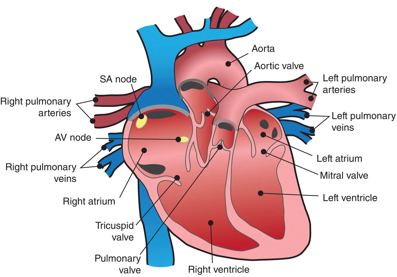
Figure 1.1 Cross section of the heart showing the chambers.
The heart is a muscle and consists of four distinct layers. The endocardium is the innermost layer and composes a lining for the interior of the heart. The epicardium is the outer layer of the heart. Between the endocardium and epicardium lies the myocardium—the thickest layer—which is muscle. The entire heart is encased in a liquid-filled sac called the pericardium, which acts like a shock absorber for the heart. The pericardial sac contains about 15–20 cc of pericardial fluid in a healthy individual. In the event that fluid builds up to abnormally high levels in the pericardial sac (such as might occur when a lead or catheter inside the heart perforates the endocardium, myocardium, and epicardium and goes exterior to the heart), this fluid can place pressure on the heart in a condition known as cardiac tamponade. Since the heart is contained in a relatively small space, this pressure can compromise the heart’s ability to fill with blood and pump efficiently. During device implantation, perforation is an important concern because it can lead to cardiac tamponade. In the event that perforation results in cardiac tamponade, a needle is inserted into the pericardial sac (through the chest wall) to drain the blood. Lead perforation does not always result in cardiac tamponade, but it is a serious concern.
Blood flow through the heart
The heart is a pump and it is located amid a network of vessels that carry deoxygenated blood into the right side of the heart and reoxygenated blood into the left side of the heart. The flow is actually fairly simple. Deoxygenated blood enters the right side of the heart and is pumped over to the lungs via the pulmonary arteries and is returned back (as oxygen-rich blood) to the left side of the heart by way of the pulmonary veins (PV). While both right and left sides of the heart contract at the same time as a single unit, the right side is busy pumping deoxygenated blood to the lungs, while the left side is pumping reoxygenated blood out to the rest of the body.
Deoxygenated blood enters the right side of the heart via the superior vena cava (SVC), but once it has become oxygenated again, blood is pumped back out from the left side of the heart into the aorta. The aorta is the largest vessel in the body, and it forms a U shape at the top of the heart. These portions of the aorta are called the ascending, the descending, and the arch. Coming off the aortic arch are three main arteries: the left subclavian artery, the left common carotid artery, and the brachiocephalic trunk.
To better understand the blood flow through the heart, it is important to review the structure of the heart. The atria or upper chambers of the heart are smaller, have thinner walls, and are smoother on the inside than the ventricles. Within the ventricles is a network of fibrous strands known as trabeculae. These structural differences become important in lead implantation within the heart; it is much easier to affix or lodge a lead in the trabeculae of the ventricles than to try to anchor the lead to a smooth atrial wall. Historically, atrial leads have almost always been active-fixation screw-in-type leads, while ventricular leads were almost always passive-fixation leads (fins or tines that lodge in the trabeculae). Today, active-fixation leads are often used in both chambers since they facilitate lead removal (Figure 1.2).
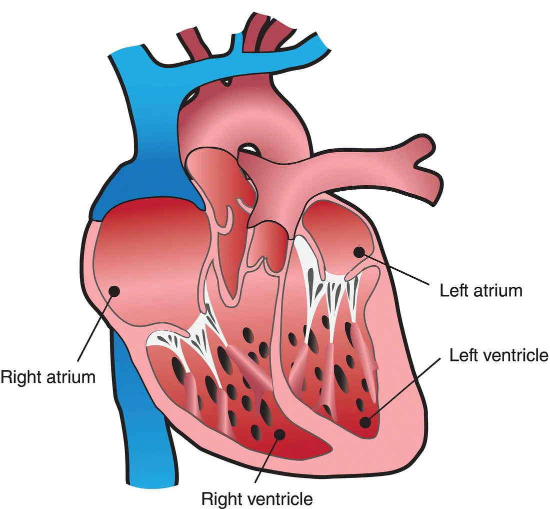
Figure 1.2 Note that the atria are smooth walled, while the ventricles contain a spongelike fibrous network of trabeculae.
Overall, blood flow to the heart is discussed, right and left sides, although it is important to recognize that what happens in the heart, that is, systole (contraction) and diastole (relaxation), are happening on both sides at the same time. The right atrium of the heart receives blood from the SVC, the inferior vena cava (IVC), and the coronary sinus (CS). The CS is technically a vein and it has an opening or ostium (sometimes just called os) at the base of the right atrium, slightly posterior. The CS delivers oxygen-depleted blood to the right atrium from the coronary arteries that encircle the exterior of the heart. The CS is of interest in cardiac resynchronization therapy (CRT) because the left ventricular lead is passed through the CS (counter to the flow of blood) in order to be placed into the coronary vessels to pace the left ventricle. CRT is used in patients with heart failure, whose hearts have remodeled,...
Table of contents
- Cover
- Title page
- Copyright page
- Preface
- Acknowledgments
- Chapter 1: Cardiovascular anatomy and physiology
- Chapter 2: Cardiac conduction system
- Chapter 3: The cardiac cycle and hemodynamics
- Chapter 4: Heart disease
- Chapter 5: Cardiac medications related to cardiac rhythm management devices
- Chapter 6: The basics of ECG and rhythm interpretation
- Chapter 7: Arrhythmia analysis
- Chapter 8: Electricity 101
- Chapter 9: Pacing 101
- Chapter 10: Indications for pacing
- Chapter 11: Pacemaker implantation
- Chapter 12: Connecting the leads to the pulse generator
- Chapter 13: Pacemaker modes and codes
- Chapter 14: Single-chamber timing cycles
- Chapter 15: Introduction to dual-chamber timing cycles
- Chapter 16: Dual-chamber timing cycles: the atrial channel
- Chapter 17: Dual-chamber timing cycles
- Chapter 18: Paced ECG and EGM analysis
- Chapter 19: Upper-rate behavior
- Chapter 20: Advanced dual-chamber timing
- Chapter 21: Rate-responsive pacing
- Chapter 22: Special features
- Chapter 23: Automatic capture algorithms
- Chapter 24: Pacemaker follow-up
- Chapter 25: Follow-up and troubleshooting
- Answer key
- Index
- End User License Agreement
Frequently asked questions
Yes, you can cancel anytime from the Subscription tab in your account settings on the Perlego website. Your subscription will stay active until the end of your current billing period. Learn how to cancel your subscription
No, books cannot be downloaded as external files, such as PDFs, for use outside of Perlego. However, you can download books within the Perlego app for offline reading on mobile or tablet. Learn how to download books offline
Perlego offers two plans: Essential and Complete
- Essential is ideal for learners and professionals who enjoy exploring a wide range of subjects. Access the Essential Library with 800,000+ trusted titles and best-sellers across business, personal growth, and the humanities. Includes unlimited reading time and Standard Read Aloud voice.
- Complete: Perfect for advanced learners and researchers needing full, unrestricted access. Unlock 1.5M+ books across hundreds of subjects, including academic and specialized titles. The Complete Plan also includes advanced features like Premium Read Aloud and Research Assistant.
We are an online textbook subscription service, where you can get access to an entire online library for less than the price of a single book per month. With over 1.5 million books across 990+ topics, we’ve got you covered! Learn about our mission
Look out for the read-aloud symbol on your next book to see if you can listen to it. The read-aloud tool reads text aloud for you, highlighting the text as it is being read. You can pause it, speed it up and slow it down. Learn more about Read Aloud
Yes! You can use the Perlego app on both iOS and Android devices to read anytime, anywhere — even offline. Perfect for commutes or when you’re on the go.
Please note we cannot support devices running on iOS 13 and Android 7 or earlier. Learn more about using the app
Please note we cannot support devices running on iOS 13 and Android 7 or earlier. Learn more about using the app
Yes, you can access The Nuts and Bolts of Implantable Device Therapy by Tom Kenny in PDF and/or ePUB format, as well as other popular books in Medicine & Cardiology. We have over 1.5 million books available in our catalogue for you to explore.