Morphology of Blood Disorders, 2nd edition is an outstanding atlas with over 800 high-quality digital images, covering the whole spectrum of blood and bone marrow morphology, with particular emphasis on malignant haematology. Originally written in the Italian language by two world leaders in the field, the book has been expertly translated by the renowned haematologist and teacher, Barbara Bain.
This book explores the major topics of haematological pathology, blending classical teaching with up-to-date WHO classification and terminology. Each image in this book is derived from material obtained for diagnostic purposes from patients with serious haematological conditions. Morphological details are supplemented by detailed descriptions of the output and role of automated instruments in disorders of the blood.
Morphology of Blood Disorders, 2nd edition is an essential reference source for diagnosis in the haematology laboratory, designed to be the go-to guide for anyone with an interest in blood cell morphology.
Trusted by 375,005 students
Access to over 1.5 million titles for a fair monthly price.
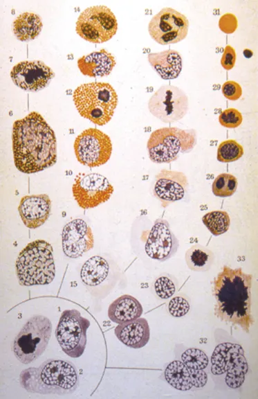
In the second half of the 19th century, developments in microscopy and in staining techniques permitted the observation of various types of blood cell, leading to a developing understanding of their heterogeneity, morphological characteristics and manner of proliferating and differentiating. The German pathologist, Neumann, recognised that the formation of blood cells from a single progenitor cell took place in the bone marrow. From this recognition, together with the work of the Russian scientist, Maximov, evolved the monophyletic or unitary theory of haemopoiesis, of which Adolfo Ferrata became one of the most convinced supporters (Fig. 1.1).1 It was the studies of McCulloch and Till, at the beginning of the 1960s, that provided the necessary experimental evidence with their demonstration of the capacity of bone marrow cells to form multilineage myeloid colonies in the spleen when injected into animals rendered aplastic by irradiation.2 This experiment was repeated in man with the first transplants of bone marrow between twins and into workers accidentally exposed to nuclear irradiation.3
Fig. 1.1 Morphogenesis of blood cells, according to the monophyletic theory, illustrated in the original table in Adolfo Ferrata's text, Le Emopatie, published in 1933 by the Società Editrice Libraria. From the morphologically undifferentiated haemopoietic progenitors, grouped together at bottom left within an incomplete circle, originate the various lineages of bone marrow precursors, which progressively develop their distinctive morphological characteristics, finally giving rise to the mature cells of the peripheral blood. From left to right the lineages are basophil granulocytes, eosinophil granulocytes, neutrophil granulocytes, erythroid series and, bottom right, megakaryocytes. The illustration is incomplete, lacking the lymphocytes and monocytes; it nevertheless shows the high level of intuition and knowledge achieved by morphological observation alone.
The blood, understood as both the haemopoietic matrix and the circulating cells, is a complicated tissue in which numerous elements are selected, organized and regulated like the instruments of a symphonic orchestra, to provide a harmonious, stable and effective result. The formation of the blood depends on the existence of a multipotent stem cell, recognised by its capacity to establish long-term cultures in vitro. This cell is at the top of a tree with increasing numbers of branches towards the base. The initial division is into two progenitors that differ in their potential, restricted respectively to lymphopoiesis and myelopoiesis. Successive branches give rise to other progenitors, organised in a hierarchy, with the capacity for differentiation becoming more limited and specific. This process culminates in the production of the population of morphologically recognisable haemopoietic cells of the bone marrow and ultimately of the mature elements that circulate in the peripheral blood.
Stem Cells
Haemopoietic stem cells are multipotent and it is from them that all the circulating blood cells derive. Haemopoiesis is the well-organised and carefully controlled process by which this fundamental capacity is realised. The adjective ‘stem’ derives from the Latin stamen, meaning thread, to which, figuratively speaking, is bound the life of every man: the thread in classical mythology is spun and then cut off by the Fates.4 The stamen in botany, moreover, is the flower's male organ. While this concept was used in the 19th century in Darwin's evolutionary theory to indicate the unicellular ancestor of all living creatures, it was only at the beginning of the 20th century that the term ‘stem cell’ was attributed to the common precursor of all the cells of the blood.5
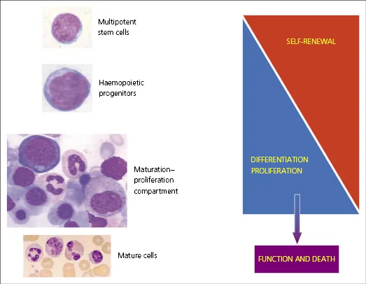
The haemopoietic stem cells have two essential properties (Fig. 1.2):
They are multipotent, that is capable of differentiating into all the various types of blood cell; this differentiation is associated with the loss of multilineage potential and the formation of committed haemopoietic progenitors;
They are capable of indefinite self-renewal, maintaining themselves through many generations as a small constant percentage of daughter cells with their multilineage potential conserved.
Fig. 1.2 Schematic diagram of the haemopoietic compartments and their principal functional characteristics. Cells of the multipotent stem cell compartment have the maximum self-renewal capacity, proliferate quite slowly and only show differentiation to a minimal extent. Cells of the compartment of haemopoietic progenitors, while maintaining the morphological uniformity of the undifferentiated cells, are more heterogeneous and organised in a hierarchical manner (see Fig. 1.6): the capacity for self-renewal is reduced, while the cells proliferate and differentiate. The cells of the maturing-proliferating compartment, which correspond to the morphologically recognisable multilineage haemopoietic cells of the bone marrow, have lost the capacity for self-renewal. From this compartment originate the mature cells that pass into the peripheral blood: these are destined to carry out their functions without dividing again (with the exception of the lymphocytes).
The maintenance of stem cell numbers is necessary to maintain effective multilineage haemopoiesis for the life of an individual. In stable conditions (homeostasis), the majority of the rare mitoses seen in stem cells are symmetrical, producing two identical daughter cells, which are destined to give rise to differentiated progeny. The maintenance of a stable number of stem cells is achieved by the simultaneous occurrence of a more limited number of asymmetric divisions, in which a single stem cell produces two non-identical daughter cells, one of which maintains the original multipotent capacity while the other takes the path of differentiation. This asymmetric mitosis could be predetermined at the moment of mitosis, as is suggested by the polarized distribution of regulatory molecules,6 messenger RNA and individual proteins, which are not distributed equally between the daughter cells. In other instances, the factors determining that one of the daughter cells takes the pathway of differentiation could reside in the microenvironment.7
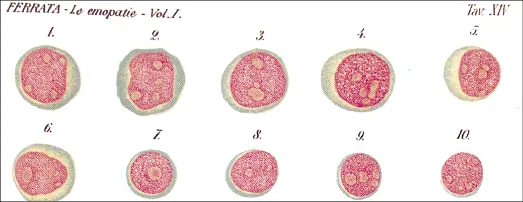
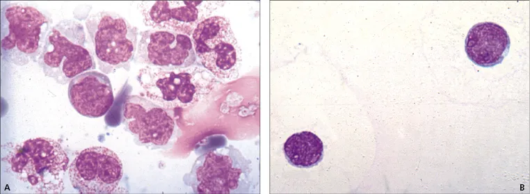
The multipotent long-term culture initiating haemopoietic stem cells (LT-HSC) can be identified and enriched in the mouse thanks to their surface membrane immunophenotype, characterised by positivity for c-Kit, Thy-1.1 (weak) and Sca-1 (stem cell antigen 1), and negativity for all the lineage-specific markers expressed by committed progenitors, differentiated precursors and mature cells. There is neither a single marker, nor a combination of markers, that is specific for haemopoietic stem cells in man. The immunophenotypic markers most often used, particularly for counting stem cells in relation to transplantation, are CD34, CD133, CD90 and CD117. The functional capacity of stem cells can be demonstrated by culture, i.e. they can give rise to long-term cultures in vitro. The morphology of these forms is similar to that of small blast cells, with a very high nucleocytoplasmic (N:C) ratio, scanty weakly basophilic cytoplasm and homogeneous nuclear chromatin, which is fairly compact (Figs 1.3, 1.4 and 1.5). In some cells the cytoplasm is more abundant and basophilic. The nucleus can have visible nucleoli of variable prominence.
Fig. 1.3 The haemocytoblasts of Ferrata (original illustration from Ferrata A, Le emopatie, 1933). The cells numbered from 8 to 10 particularly evoke the morphology and size of stem cells and haemopoietic progenitors as one recognises them today (see Figs 1.4 and 1.5). The other cells are more similar to myeloblasts, with an angulated nuclear outline (e.g. 1 and 2), or lymphoblasts.
Fig. 1.4 Morphology of haemopoietic stem cells. (A) Cytocentrifuge preparation of mononuclear cells harvested for pe...
CHAPTER 8: Acute Lymphoblastic and Mixed Phenotype Leukaemia
CHAPTER 9: Neoplasms of Mature B, T and NK Cells
CHAPTER 10: Plasma Cell Proliferative Disorders
CHAPTER 11: Reactive Bone Marrow Changes and Non-haemopoietic Neoplasms
Index
End User License Agreement
Frequently asked questions
Yes, you can cancel anytime from the Subscription tab in your account settings on the Perlego website. Your subscription will stay active until the end of your current billing period. Learn how to cancel your subscription
No, books cannot be downloaded as external files, such as PDFs, for use outside of Perlego. However, you can download books within the Perlego app for offline reading on mobile or tablet. Learn how to download books offline
Perlego offers two plans: Essential and Complete
Essential is ideal for learners and professionals who enjoy exploring a wide range of subjects. Access the Essential Library with 800,000+ trusted titles and best-sellers across business, personal growth, and the humanities. Includes unlimited reading time and Standard Read Aloud voice.
Complete: Perfect for advanced learners and researchers needing full, unrestricted access. Unlock 1.5M+ books across hundreds of subjects, including academic and specialized titles. The Complete Plan also includes advanced features like Premium Read Aloud and Research Assistant.
Both plans are available with monthly, semester, or annual billing cycles.
We are an online textbook subscription service, where you can get access to an entire online library for less than the price of a single book per month. With over 1.5 million books across 990+ topics, we’ve got you covered! Learn about our mission
Look out for the read-aloud symbol on your next book to see if you can listen to it. The read-aloud tool reads text aloud for you, highlighting the text as it is being read. You can pause it, speed it up and slow it down. Learn more about Read Aloud
Yes! You can use the Perlego app on both iOS and Android devices to read anytime, anywhere — even offline. Perfect for commutes or when you’re on the go. Please note we cannot support devices running on iOS 13 and Android 7 or earlier. Learn more about using the app
Yes, you can access Morphology of Blood Disorders by Giuseppe d'Onofrio,Gina Zini, Barbara J. Bain in PDF and/or ePUB format, as well as other popular books in Medicine & Hematology. We have over 1.5 million books available in our catalogue for you to explore.