eBook - ePub
Surface Electromyography
Physiology, Engineering, and Applications
- English
- ePUB (mobile friendly)
- Available on iOS & Android
eBook - ePub
Surface Electromyography
Physiology, Engineering, and Applications
About this book
Reflects on developments in noninvasive electromyography, and includes advances and applications in signal detection, processing and interpretation
- Addresses EMG imaging technology together with the issue of decomposition of surface EMG
- Includes advanced single and multi-channel techniques for information extraction from surface EMG signals
- Presents the analysis and information extraction of surface EMG at various scales, from motor units to the concept of muscle synergies.
Tools to learn more effectively

Saving Books

Keyword Search

Annotating Text

Listen to it instead
Information
1
Physiology of Muscle Activation and Force Generation
R. M. Enoka1 and J. Duchateau2
1Department of Integrative Physiology, University of Colorado, Boulder, Colorado
2Laboratory of Applied Biology and Neurophysiology, ULB Neuroscience Institute (UNI), Université Libre de Bruxelles (ULB), Brussels, Belgium
1.1 Introduction
To extract information about the control of movement by the nervous system from electromyographic (EMG) signals, it is necessary to understand the processes underlying both the generation of the activation signal and the torques exerted by the involved muscles. As a foundation for the subsequent chapters in this book, the goal of this chapter is to describe the physiology of muscle activation and force generation. We discuss the anatomy of the final common pathway from the nervous system to muscle, the electrical properties of motor neurons and muscle fibers, the contractile properties of muscle fibers and motor units, the concept of motor unit types, and the control of muscle force by modulating the recruitment and rate coding of motor unit activity.
1.2 Anatomy of a Motor Unit
The basic functional unit of the neuromuscular system is the motor unit. It comprises a motor neuron, including its dendrites and axon, and the muscle fibers innervated by the axon [28]. The motor neuron is located in the ventral horn of the spinal cord or brain stem where it receives sensory and descending inputs from other parts of the nervous system. The axon of each motor neuron exits the spinal cord through the ventral root, or through a cranial nerve in the brain stem, and projects in a peripheral nerve to its target muscle and the muscle fibers it innervates. Because the generation of an action potential by a motor neuron typically results in the generation of action potentials in all of the muscle fibers belonging to the motor unit, EMG recordings of muscle fiber action potentials provide information about the activation of motor neurons in the spinal cord or brain stem.
1.2.1 Motor Nucleus
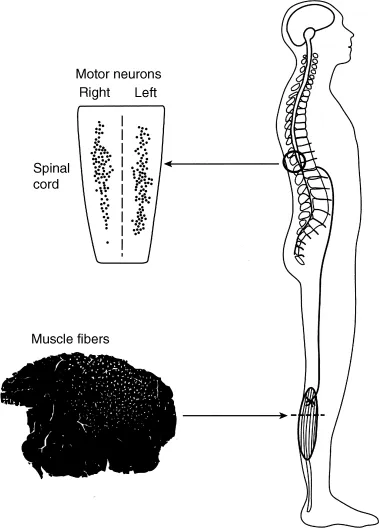
The population of motor neurons that innervate a single muscle is known as a motor nucleus or motor neuron pool [51]. The number of motor neurons in a motor nucleus ranges from a few tens to several hundred [40,58] (Table 1.1). The motor neuron pool for each muscle typically extends longitudinally for a few segments of the spinal cord (Fig. 1.1), and at each segmental level the pools for proximal muscles tend to be more ventral and lateral than those for distal muscles and the pools for anterior muscles are more lateral than those for posterior muscles [59]. Nonetheless, the extensive dendritic projections of motor neurons intermingle across motor neuron pools.
Table 1.1 Motor Neuron Locations and Numbers for Selected Forelimb Muscles
| Muscle | Spinal Location | Number |
| Biceps brachii | C5–C7 | 1051 |
| Triceps brachii | C6–T1 | 1271 |
| Flexor carpi radialis | C7–C8 | 235 |
| Extensor carpi radialis | C5–C7 | 890 |
| Flexor carpi ulnaris | C7–T1 | 314 |
| Extensor carpi ulnaris | C7–T1 | 216 |
| Extensor pollicis longus | C8–T1 | 14 |
| Abductor pollicis longus | C8–T1 | 126 |
| Flexor digitorum superficialis | C8–T1 | 306 |
| Extensor digitorum communis | C8–T1 | 273 |
| Flexor digitorum profundus | C8–T1 | 475 |
| Extensor digiti secundi proprius | C8–T1 | 87 |
| Abductor pollicis brevis and flexor pollicis brevis | C8–T1 | 115 |
| Adductor pollicis | C8–T1 | 370 |
| First dorsal interosseus | C8–T1 | 172 |
| Lateral lumbricalis | C8–T1 | 57 |
| Data are from Jenny and Inukai [58] and are listed as pairs of antagonistic muscles. | ||

Figure 1.1 Muscle force is controlled by a population of motor units (motor unit pool) located in the spinal cord with each motor unit innervating a number of muscle fibers (muscle unit). The muscle fibers belonging to a single muscle unit are indicated by the white dots in the cross-sectional view of the muscle. A typical motor unit pool spans several spinal segments, and muscle units are usually limited to discrete parts of the muscle. Modified from Enoka [30] with permission.
1.2.2 Muscle Fibers
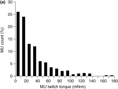
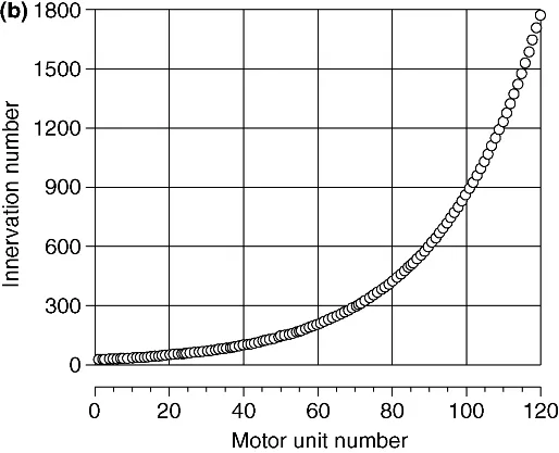
The muscle fibers innervated by a single axon are known as the muscle unit (Fig. 1.1), the size of which varies across each motor unit pool. The motor units first recruited during a voluntary contraction innervate fewer muscle fibers and hence have smaller muscle units than those that are recruited later in the contraction. Most motor units in a muscle have small muscle units and only a few have large muscle units [76,102,107] (Fig. 1.2A). Based on the association between muscle unit size and maximal motor unit force, Enoka and Fuglevand [32] estimated the innervation numbers (muscle unit size) for the 120 motor units in a human hand muscle (first dorsal interosseus) ranged from 21 to 1770 (Fig. 1.2B). Similar relations likely exist for most muscles [51]. Due to the exponential distribution of innervation number across a motor unit pool, it is necessary to distinguish between the number of motor unit action potentials discharged from the spinal cord and the number of muscle fiber action potentials recorded in the muscle with EMG electrodes. This distinction is indicated with the term “neural drive” to denote the motor unit action potentials and “muscle activation” to indicate the muscle fiber action potentials [26,31,36].


Figure 1.2 Variation in muscle unit size across the motor unit pool. (a) Distribution of motor unit (MU) twitch torques for 528 motor units in the tibialis anterior muscle of 10 subjects [107]. (b) Estimated distribution of innervation numbers across the 120 motor units comprising the first dorsal interosseus muscle [32].
The fibers in each muscle unit are located in a subvolume of the muscle and intermingle with the fibers of other muscle units (Fig. 1.1). The spatial distribution of the fibers belonging to a muscle unit is referred to as the motor unit territory. Counts of muscle unit fibers indicate that motor unit territories can occupy from 10% to 70% of the cross-sectional area of a muscle and that the density of muscle unit fibers ranges from 3 to 20 per 100 muscle fibers [51]. Moreover, the fibers of a single muscle unit often do not extend from one end of the muscle to the other, but instead terminate within a muscle fascicle [48,108]. As a consequence of muscle unit anatomy, the forces generated by individual muscle fibers must be transmitted through various layers of connective tissues before reaching the skeleton and contributing to the movement. Such interactions attenuate the unique contribution of individual fibers to the net muscle force during a movement and thereby reduce the influence of differences in contractile properties among muscle fibers.
1.3 Motor Neuron
The motor unit is classically considered to be the final common pathway in that sensory and descending inputs converge onto a single neuron that discharges an activation signal to the muscle fibers it innervates [28]. The motor neuron has extensive dendritic branches that receive up to 50,000 synaptic contacts with each contact capable of eliciting inward or outward currents across the membrane...
Table of contents
- Cover
- Series Page
- Title Page
- Copyright
- Introduction
- Acknowledgments
- Contributors
- Chapter 1: Physiology of Muscle Activation and Force Generation
- Chapter 2: Biophysics of the Generation of EMG Signals
- Chapter 3: Detection and Conditioning of Surface EMG Signals
- Chapter 4: Single-Channel Techniques for Information Extraction from the Surface EMG Signal
- Chapter 5: Techniques for Information Extraction from the Surface EMG Signal: High-Density Surface EMG
- Chapter 6: Muscle Coordination, Motor Synergies, and Primitives from Surface EMG
- Chapter 7: Surface EMG Decomposition
- Chapter 8: EMG Modeling and Simulation
- Chapter 9: Electromyography-Driven Modeling for Simulating Subject-Specific Movement at the Neuromusculoskeletal Level
- Chapter 10: Muscle Force and Myoelectric Manifestations of Muscle Fatigue in Voluntary and Electrically Elicited Contractions
- Chapter 11: EMG of Electrically Stimulated Muscles
- Chapter 12: Surface EMG Applications in Neurophysiology
- Chapter 13: Surface EMG in Ergonomics and Occupational Medicine
- Chapter 14: Applications in Proctology and Obstetrics
- Chapter 15: EMG and Posture in Its Narrowest Sense
- Chapter 16: Applications in Movement and Gait Analysis
- Chapter 17: Applications in Musculoskeletal Physical Therapy
- Chapter 18: Surface EMG Biofeedback
- Chapter 19: EMG in Exercise Physiology and Sports
- Chapter 20: Surface Electromyography for Man–Machine Interfacing in Rehabilitation Technologies
- Index
- IEEE Press Series in Biomedical Engineering
- End User License Agreement
Frequently asked questions
Yes, you can cancel anytime from the Subscription tab in your account settings on the Perlego website. Your subscription will stay active until the end of your current billing period. Learn how to cancel your subscription
No, books cannot be downloaded as external files, such as PDFs, for use outside of Perlego. However, you can download books within the Perlego app for offline reading on mobile or tablet. Learn how to download books offline
Perlego offers two plans: Essential and Complete
- Essential is ideal for learners and professionals who enjoy exploring a wide range of subjects. Access the Essential Library with 800,000+ trusted titles and best-sellers across business, personal growth, and the humanities. Includes unlimited reading time and Standard Read Aloud voice.
- Complete: Perfect for advanced learners and researchers needing full, unrestricted access. Unlock 1.4M+ books across hundreds of subjects, including academic and specialized titles. The Complete Plan also includes advanced features like Premium Read Aloud and Research Assistant.
We are an online textbook subscription service, where you can get access to an entire online library for less than the price of a single book per month. With over 1 million books across 990+ topics, we’ve got you covered! Learn about our mission
Look out for the read-aloud symbol on your next book to see if you can listen to it. The read-aloud tool reads text aloud for you, highlighting the text as it is being read. You can pause it, speed it up and slow it down. Learn more about Read Aloud
Yes! You can use the Perlego app on both iOS and Android devices to read anytime, anywhere — even offline. Perfect for commutes or when you’re on the go.
Please note we cannot support devices running on iOS 13 and Android 7 or earlier. Learn more about using the app
Please note we cannot support devices running on iOS 13 and Android 7 or earlier. Learn more about using the app
Yes, you can access Surface Electromyography by Roberto Merletti, Dario Farina, Roberto Merletti,Dario Farina in PDF and/or ePUB format, as well as other popular books in Biological Sciences & Biotechnology. We have over one million books available in our catalogue for you to explore.