eBook - ePub
Shimizu's Dermatology
Hiroshi Shimizu
This is a test
Share book
- English
- ePUB (mobile friendly)
- Available on iOS & Android
eBook - ePub
Shimizu's Dermatology
Hiroshi Shimizu
Book details
Book preview
Table of contents
Citations
About This Book
Shimizu's Dermatology, Second Edition provides practical, didactic, and rapid-reference advice on diagnosis and management of the most common dermatologic conditions. Written by one of the world's leading experts, and a best-seller in Japan where it was first published, the second edition is cohesive, richly illustrated, attractively presented, and fully revised to reflect the latest in clinical developments. This complete dermatology resource offers:
- Over 2, 000 clinical images to aid rapid and easy diagnosis
- 100% clinically focused chapters describing the clinical features, classifications, pathogeneses, pathologies, treatments and lab findings of diseases
- Key points and tips and tricks in every chapter for practical guidance
- Attractive color presentation throughout, with high-quality clinical images
- Improve your knowledge of skin conditions and their management with this concise, user-friendly, alternative to larger reference books.
Shimizu's Dermatology is ideal for specialists in clinical practice, trainees managing patients at clinics or hospitals or preparing for board exams, and medical students.
Frequently asked questions
How do I cancel my subscription?
Can/how do I download books?
At the moment all of our mobile-responsive ePub books are available to download via the app. Most of our PDFs are also available to download and we're working on making the final remaining ones downloadable now. Learn more here.
What is the difference between the pricing plans?
Both plans give you full access to the library and all of Perlego’s features. The only differences are the price and subscription period: With the annual plan you’ll save around 30% compared to 12 months on the monthly plan.
What is Perlego?
We are an online textbook subscription service, where you can get access to an entire online library for less than the price of a single book per month. With over 1 million books across 1000+ topics, we’ve got you covered! Learn more here.
Do you support text-to-speech?
Look out for the read-aloud symbol on your next book to see if you can listen to it. The read-aloud tool reads text aloud for you, highlighting the text as it is being read. You can pause it, speed it up and slow it down. Learn more here.
Is Shimizu's Dermatology an online PDF/ePUB?
Yes, you can access Shimizu's Dermatology by Hiroshi Shimizu in PDF and/or ePUB format, as well as other popular books in Medicina & Dermatología. We have over one million books available in our catalogue for you to explore.
Information
CHAPTER 1
Structure and function of the skin
The skin is the largest organ of the human body, covering a surface area of approximately 1.6 m2 and accounting for about 16% of an adult’s body weight. The skin is in direct contact with the outside environment and helps maintain four key functions:
- retention of water and other molecules
- regulation of body temperature
- protection of the body from invading microorganisms and other harmful external factors
- sensory perception.
The horny cell layer plays a crucial role in maintaining the human body’s water balance. To gain insight into cutaneous biology and skin diseases, an understanding of the structure and functions of normal human skin is essential. This chapter discusses the structure and function of normal skin and skin appendages, and the basics of immune mechanisms that mainly relate to the skin.
Overview of the skin
The skin is the largest human organ in both area and weight. It has the important function of separating the body from the external environment and maintaining homeostasis in the body. In order to achieve this, the skin has a complicated structure and various functions. Skin consists of a three‐layer structure of epidermis, dermis, and subcutaneous tissue (Fig. 1.1). The cells in the epidermis are keratinocytes (~95%), melanocytes (~5%), and Langerhans cells. The horny cell layer, formed by the keratinization of keratinocytes, is the outermost skin layer and is in direct contact with the external environment. It has the important functions of moisture retention and protection against the external environment, such as foreign substances and ultraviolet radiation. The major dermal components are collagen fibers, elastic fibers, blood vessels, and the components that maintain the skin, such as the extracellular matrix. The dermal appendages include the sweat glands, sebaceous glands, and hair follicles. At the boundary between the epidermis and dermis are finger‐like projections, called dermal papillae, that project into the epidermis (see Fig. 1.1). The epidermis also projects into the dermis, producing the epidermal rete ridge. The main component of subcutaneous tissue is fat tissue, which stores neutral fat and provides protection from external impacts and heat/cold.

Fig. 1.1 Structure of the skin.
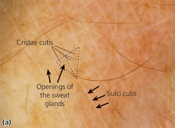
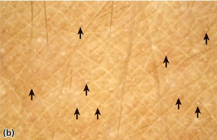
The skin surface is not smooth; it is laced with networks of fine shallow and deep grooves called sulci cutis. Sweat ducts open onto the skin surface in the center of the crista cutis, which is formed by the borders of the sulci cutis (Fig. 1.2).


Fig. 1.2 Appearance of the skin surface. a: Cristae cutis (triangle), sulci cutis and openings of the sweat glands. b: Sweat pores fed by sweat glands open to the cristae cutis (arrows).
The orientation of the sulci cutis is site dependent and is called the dermal ridge pattern. Fingerprints and patterns on the palms and soles, which are unique to each person, are formed by the sulci cutis. Langer's lines, also called cleavage lines, is a term used to define the direction within the human skin along which the skin has the least flexibility (Fig. 1.3). Elastic fibers are aligned in specific site‐dependent orientations in the dermis. Several skin diseases, such as epidermal nevi, occur along specific lines called Blaschko’s lines. These lines are distributed a...