Signaling Pathways in Liver Diseases
  1. English
  2. ePUB (mobile friendly)
  3. Available on iOS & Android
eBook - ePub

About this book

Signaling Pathways in Liver Diseases, Third Edition again provides hepatologists and hepatology researchers with an expert overview of the complex and novel cellular/extracellular signaling pathways in the liver, and their role in liver diseases. The last few years have seen a great number of developments in this field, which in turn have led to new opportunities for innovative treatments; however, the intricacy of these pathways and their interactions continue to provide a real challenge for clinicians. This outstanding book compiles the emerging knowledge into a single expert resource, cataloguing and organizing it into an accessible and understandable format.

With increased focus on the comprehension of cellular mechanisms involved in steatohepatitis, cirrhosis, and liver tumors, which has led to changes in the management of these diseases, this new edition also sees the introduction of exciting new chapters on key emerging areas such as:

  • Autophagy
  • Notch Pathway
  • P13K/PTEN Signaling in Liver Diseases
  • Sirtuins
  • Hepcidin and Iron
  • Epigenetic Regulation of Hepatic Stellate Cells and Liver Fibrosis
  • Oxidative Stress and Signaling in the Liver.

Professors Dufour and Clavien have assembled an all-star cast of chapter authors, each of whom has provided clear and appropriate illustrations to reinforce the text, with a key points box offering a concise and handy summary. Self-assessment questions and answers allow the reader to test their own knowledge.

Signaling Pathways in Liver Disease, Third Edition is the perfect educational and reference tool to bridge the information exchange between the laboratory, the clinical ward, and the operating room, and an essential tool for the modern-day hepatologist.

Frequently asked questions

Yes, you can cancel anytime from the Subscription tab in your account settings on the Perlego website. Your subscription will stay active until the end of your current billing period. Learn how to cancel your subscription.
At the moment all of our mobile-responsive ePub books are available to download via the app. Most of our PDFs are also available to download and we're working on making the final remaining ones downloadable now. Learn more here.
Perlego offers two plans: Essential and Complete
  • Essential is ideal for learners and professionals who enjoy exploring a wide range of subjects. Access the Essential Library with 800,000+ trusted titles and best-sellers across business, personal growth, and the humanities. Includes unlimited reading time and Standard Read Aloud voice.
  • Complete: Perfect for advanced learners and researchers needing full, unrestricted access. Unlock 1.4M+ books across hundreds of subjects, including academic and specialized titles. The Complete Plan also includes advanced features like Premium Read Aloud and Research Assistant.
Both plans are available with monthly, semester, or annual billing cycles.
We are an online textbook subscription service, where you can get access to an entire online library for less than the price of a single book per month. With over 1 million books across 1000+ topics, we’ve got you covered! Learn more here.
Look out for the read-aloud symbol on your next book to see if you can listen to it. The read-aloud tool reads text aloud for you, highlighting the text as it is being read. You can pause it, speed it up and slow it down. Learn more here.
Yes! You can use the Perlego app on both iOS or Android devices to read anytime, anywhere — even offline. Perfect for commutes or when you’re on the go.
Please note we cannot support devices running on iOS 13 and Android 7 or earlier. Learn more about using the app.
Yes, you can access Signaling Pathways in Liver Diseases by Jean-Francois Dufour, Pierre-Alain Clavien, Jean-Francois Dufour,Pierre-Alain Clavien in PDF and/or ePUB format, as well as other popular books in Medicine & Gastroenterology & Hepatology. We have over one million books available in our catalogue for you to explore.

Information

CHAPTER 1
Hepatocytes

Jean-François Dufour1 and Joachim C. Mertens2
1 University Clinic for Visceral Surgery and Medicine, Inselspital, University of Berne, Berne, Switzerland
2 Division of Gastroenterology and Hepatology, University Hospital Zurich, Zurich, Switzerland

KEY POINTS

  • The liver is the largest metabolic organ of the human body with a multitude of physiological functions.
  • Hepatocytes differentiate from cells of the anterior entoderm under the influence of fibroblast growth factors and bone morphogenic proteins.
  • The hepatocytes are the central parenchymal cells, performing all important biochemical processes in close contact with the bloodstream, forming a network of bile canaliculi with their cell membranes that excrete biliary fluid and its various contents into the small bowel.
  • The cell membranes of the hepatocyte show distinct domains equipped with specific transporters essential for the uptake of molecules from the bloodstream and on the opposite side of the cell for secretion into the bile.
  • The hepatocyte is an important storage compartment for carbohydrates and lipids, the main site of amino acid metabolism and protein synthesis (i.e. serum albumin, transport proteins and many coagulation factors) as well as the site of the two stages of biotransformation facilitating excretion of many toxins and xenobiotics.

Introduction

The liver, located in the right upper abdomen, is the largest metabolic organ of the human body with an average weight of 1500 g. It is an organ with a multitude of physiological functions. The complex physiological tasks the liver fulfills in the organism can be divided into three basic categories.
  • Resorption and storage
  • Synthesis and secretion
  • Detoxification and excretion
In the liver tissue that consists of a large array of diverse cells, the hepatocyte is the pivotal parenchymal cell for all physiological liver functions and by far the most abundant cell population, accounting for approximately 80% of the cytoplasmic mass of the liver [1].
The organ tissue architecture in many ways reflects its function. The liver receives blood inflow from two circulations: an arterial blood supply through the proper hepatic artery and as a distinctive feature, a second inflow of venous blood from most intraabominal organs via the portal vein. In particular, the venous blood from the gut, pancreas and spleen is drained into the portal circulation of the liver. It is this portal venous blood and its content of nutrients, toxins and xenobiotics that feeds into the various liver functions ranging from nutrient resorption and storage, detoxification and excretion of xenobiotics to biomolecule synthesis from resorbed nutrients and secretion of hormones into the bloodstream. The blood leaves the liver through the hepatic veins towards the heart. In addition to the vasculature, the liver possesses an important additional system of vessels, the arborescent system of bile ducts which transport the bile fluid produced by the hepatocytes out of the liver into the small bowel.
The further macro- and microarchitecture of the liver is determined by the aforementioned vessels and ducts with their surrounding hepatocytes and stromal cells. Along these anatomical structures the liver tissue can be divided into different functional units (Figure 1.1). The classic anatomical liver lobule has a hexagonal outline and is defined by a single terminal branch of the hepatic vein in its center with a bile duct, a terminal branch of the hepatic artery (arteriole) and a terminal branch of the portal vein (venule) on each of the six corners. This triad of portal venule, arteriole and bile duct is also called the ‘‘portal triad.” In contrast to the classic lobule, the so-called portal lobule is centered around this portal triad and has a triangular shape with the corners defined by the adjacent three portal veins. Besides these two lobules, the third and most functionally relevant unit that can be defined is the ‘‘liver acinus.” It expands between two portal triads and the neighboring two central veins and has a diamond shape. The liver acinus best reflects the metabolic functions. It is traditionally divided into three zones with the zone I or the periportal zone being closest to the arterial vascular supply of the portal triad and receiving the most oxygenation. Zones II and III are closer to the central vein and consequently less well oxygenated, making them more susceptible to hypoxic damage in situations of poor oxygen supply [2]. The zones of the liver acinus not only differ in oxygenation but contain hepatocyte populations that specialize in different metabolic functions. Oxidative energy metabolism, amino acid catabolism, cholesterol metabolism and fatty acid ß-oxidation take place primarily in the periportal zone I while lipogenesis, ketogenesis and metabolism of xenobiotics are localized pericentrally in zone III.
c1-fig-0001
Figure 1.1 Functional units of the liver parenchyma. The liver lobule with its hexagonal outline (orange), the portal lobule (red triangle) centered on its portal triad and, the most metabolically active unit, the liver acinus, which expands between two portal triads and the neighboring two central veins (diamond shape) (left). Liver portal triad (right).
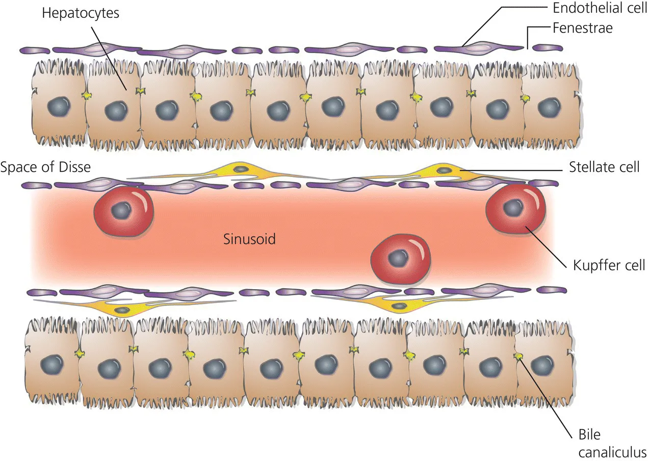
Within these anatomical structures, the hepatocytes are arranged in plates that consist of double rows of hepatocytes each flanked by a small vessel, the liver sinusoid, which contains a mixture of portal and arterial blood (Figure 1.2). The gap between the discontinuous, fenestrated endothelial cell lining of the liver sinusoids and the hepatocytes is called the perisinusoidal space or space of Disse. Hepatocyte microvilli protrude into the space of Disse and hepatic stellate cells, sometimes called Ito cells, are located in this perisinusoidal space. The adjacent hepatocyte rows form a bile channel or canaliculus running in the middle of the plate with the bile flow in the opposite direction of the inflowing sinusoidal blood, towards the bile duct of the portal triad. This tissue architecture on the one hand provides for a maximized contact surface between the hepatocytes and the sinusoidal blood and on the other the secretory surface for bile secretion.
c1-fig-0002
Figure 1.2 Hepatocytes and sinusoids. Hepatocytes are arranged in plates separated by liver blood sinusoids, which are lined by fenestrated endothelial cells. The extravascular space between the liver sinusoids and the hepatocytes is called the perisinusoidal space or space of Disse.

Liver and hepatocyte development

During organogenesis, hepatocytes derive from the anterior portion of the definitive endoderm after completion of the embryonic gastrulation [3] . Cell lineage tracing in mouse embryos shows three regions in the medial and bilateral foregut as sources of hepatic progenitor cells [4]. Upon closure of the foregut, progenitor cells from these regions come to lie in immediate proximity to the developing heart whose mesoderm has been shown to provide fibroblast growth factors 1 and 2 (FGF1 and FGF2) crucial for the induction of the hepatic cell differentiation as determined by the initiation of albumin expression [5]. Besides FGF, bone morphogenetic proteins (BMP-2 and BMP-4), similarly derived from the septum transversum mesenchyme, were found to play an important role in hepatogenesis [6]. Both FGF and BMP signaling appear to be counterbalanced by transforming growth factor-β (TGF-β) signals that prevent inappropriate differentiation and as such represent a timer of embryonic liver development [7]. The Wnt signaling pathway has also been implicated in hepatogenesis during recent years but its function appears to be more complex than the clearly instructive function of the FGF and BMP signaling, with a time- and location-dependent Wnt-mediated differential regulation of the Hhex transcription factor.
Besides the central signaling molecules, pivotal transcription factors that have been described in hepatogenesis and the induction of hepatocyte-typical gene expression are Foxa2, GATA-4, C/EBPβ and HNF1β, which can form a transcription complex inducing transcription of the albumin gene [8]. Among the earliest genes expressed in the hepatic endoderm that indicated a hepatic cell fate are Albumin, Afp, Ttr (transthyretin), Rbp (retinol binding protein), and the transcription factor Hnf4a [9].
During the further development of the hepatic bud transcription factors Hhex and Prox1 have been identified as regulators of the dynamic cellular interaction and the associated matrix metalloprotease (MMP)-dependent remodeling of the extracellular matrix [10,11].
The hepatocyte precursor cells or hepatoblasts that form the hepatic bud appear to be pluripotent with the potential to differentiate into hepatocytes or cholangiocytes. Those hepatoblasts that differentiate into hepatocytes show a further embryonic and postnatal maturation during which they develop the typical gene expression profile and phenotype of adult hepatocytes [12]. This maturation of a hepatocyte gene expression profile depends on the complex interplay of a network of transcription factors with a set of six transcription factors in its center (HNF1a, HNF1β, FoxA2, HNF4a1, HNF6, and LRH-1). This highly and, with progressing differentiation, increasingly cross-regulated transcription factor network is thought to ensure correct terminal differentiation of hepatocytes [13].
Besides the differentiation into hepatocytes, a number of hepatoblasts differentiate into cholangiocytes which then line the intrahepatic bile ducts. Expression of the transcription factor Sox9 is considered the earliest indicator of cholangiocyte differentiation. Sox9 has been demonstrated as regulator of the bile duct development and accordingly Sox9-expressing cells first appear in the vicinity of the portal venules and form the so-called ductal plate that encloses the periportal mesenchyme [14]. Ductal plate formation appears to depend on a TGF-β gradient with a critical required concentration for appropriate biliary differentiation. The Notch signaling pathway has been proposed as the second important pathway for cholangiocyte differentiation. Notch signaling seems to regulate the initial d...

Table of contents

  1. Cover
  2. Title page
  3. Table of Contents
  4. List of Contributors
  5. CHAPTER 1: Hepatocytes
  6. CHAPTER 2: Signaling pathways in biliary epithelial cells
  7. CHAPTER 3: Stellate cells
  8. CHAPTER 4: Kupffer cells
  9. CHAPTER 5: Hepatic sinusoidal endothelial cells
  10. CHAPTER 6: Extracellular matrix
  11. CHAPTER 7: Platelets: a new cell type in liver physiology
  12. CHAPTER 8: Immune cell communication in liver disease and liver regeneration
  13. CHAPTER 9: Extracellular vesicle RNA in liver disease
  14. CHAPTER 10: Endoplasmic reticulum stress in nonalcoholic fatty liver disease
  15. CHAPTER 11: Autophagy
  16. CHAPTER 12: CXC chemokine receptor signaling in liver repair and regeneration
  17. CHAPTER 13: Metabolic regulation of liver regeneration
  18. CHAPTER 14: TNF signaling
  19. CHAPTER 15: Fas/FasL
  20. CHAPTER 16: Interferon signaling
  21. CHAPTER 17: Endocannabinoid signaling in liver pathologies
  22. CHAPTER 18: The WNT/β-catenin pathway
  23. CHAPTER 19: Hedgehog signaling in the liver
  24. CHAPTER 20: Notch pathway
  25. CHAPTER 21: PI3K/PTEN signaling in liver diseases
  26. CHAPTER 22: mTOR signaling in liver disease
  27. CHAPTER 23: LKB1/AMPK pathway in the control of hepatic energy metabolism
  28. CHAPTER 24: NF-κB
  29. CHAPTER 25: c-Jun NH2-terminal kinases in liver diseases
  30. CHAPTER 26: p53
  31. CHAPTER 27: Sirtuins
  32. CHAPTER 28: Bile acids and their receptors
  33. CHAPTER 29: Hepcidin and iron
  34. CHAPTER 30: Gut microbiome and liver diseases
  35. CHAPTER 31: Epigenetic regulation of hepatic stellate cells and liver fibrosis
  36. CHAPTER 32: Signaling in hepatocellular carcinoma
  37. CHAPTER 33: Interplay between hepatitis B virus and innate immune signaling pathways
  38. CHAPTER 34: Signaling of hepatitis C virus
  39. CHAPTER 35: Oxidative stress and signaling in the liver
  40. Answers to MCQs
  41. Index
  42. End User License Agreement