Nanotechnology for Biomedical Imaging and Diagnostics: From Nanoparticle Design to Clinical Applications reflects upon the increasing role of nanomaterials in biological and medical imaging, presenting a thorough description of current research as well as future directions. With contributions from experts in nanotechnology and imaging from academia, industry, and healthcare, this book provides a comprehensive coverage of the field, ranging from the architectural design of nanomaterials to their broad imaging applications in medicine.
Grouped into three sections, the book:
Elucidates all major aspects of nanotechnology and bioimaging
Provides comprehensive coverage of the field, ranging from the architectural design of nanomaterials to their broad imaging applications in medicine
Written by well-recognized experts in academia, industry, and healthcare, will be an excellence source of reference
With a multidisciplinary approach and a balance of research and diagnostic topics, this book will appeal to students, scientiests, and healthcare professionals alike
Trusted by 375,005 students
Access to over 1.5 million titles for a fair monthly price.
1 Historical Perspective on Nanoparticles in Imaging from 1895 to 2000
Mikhail Y. Berezin
Department of Radiology, Washington University School of Medicine, St. Louis, MO, USA
1.1 Introduction
Out of the two main subjects covered in this book—imaging and technology—imaging, or more commonly referred to as radiology, “the eye of medicine,” is certainly the oldest. Prior to the appearance of nanoscience, radiology had already been well established through several generations of physicians who themselves processed thousands of images every year. Still, the persistent quest to “see the invisible” to better diagnose patients forced radiologists to pay close attention to the research and development of new imaging technologies. In the past two decades, nanoparticle contrast agents, stemming from the earliest contrast agents discovered soon after the discovery of X-rays over a hundred years ago, have become the holy grail of imaging. Today, an impressive number of radiological procedures that routinely utilize nanoparticles in clinics with even more impressive number are under preclinical testing and medical research.
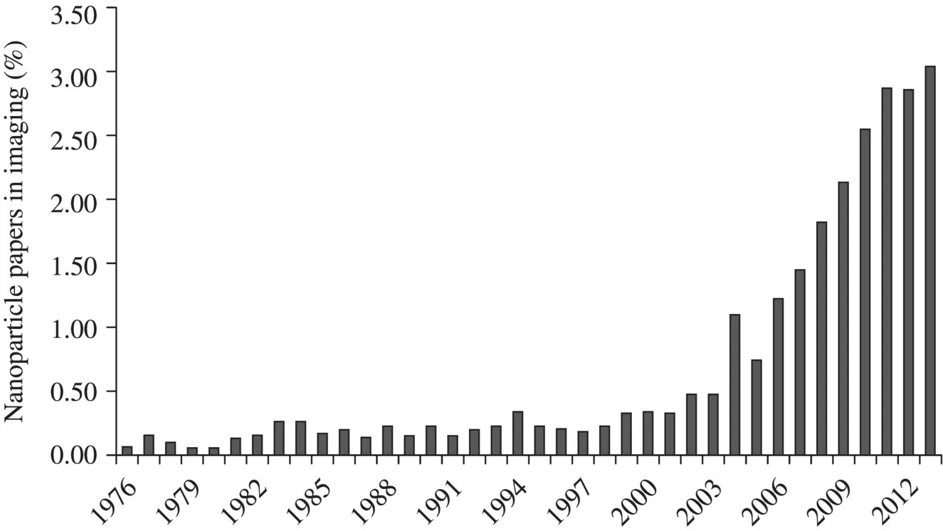
The National Institutes of Health (NIH) in 2002 prioritized the most pressing problems facing medical science and identified three key areas in need of research: biological pathways, molecular imaging, and nanotechnology. The focus on these three critical components, backed by substantial investments from the NIH, transformed classic radiology and early disorchestrated attempts with nanoparticles into a mature field known today as molecular imaging. Figure 1.1 reflects a remarkable tenfold increase in nanoparticle-related medical imaging research from a relatively modest approximately 0.25–0.3% in the twentieth century to the current 3%. This growth resulted in more than 1500 nanoparticle imaging-related publications in 2012 alone.
Figure 1.1 Growth of the nanoparticle research in biomedical imaging. Solid arrows show the appearance of imaging techniques, and dotted arrows show the emergence of nanoparticles. A number of citations are given from PubMed database.
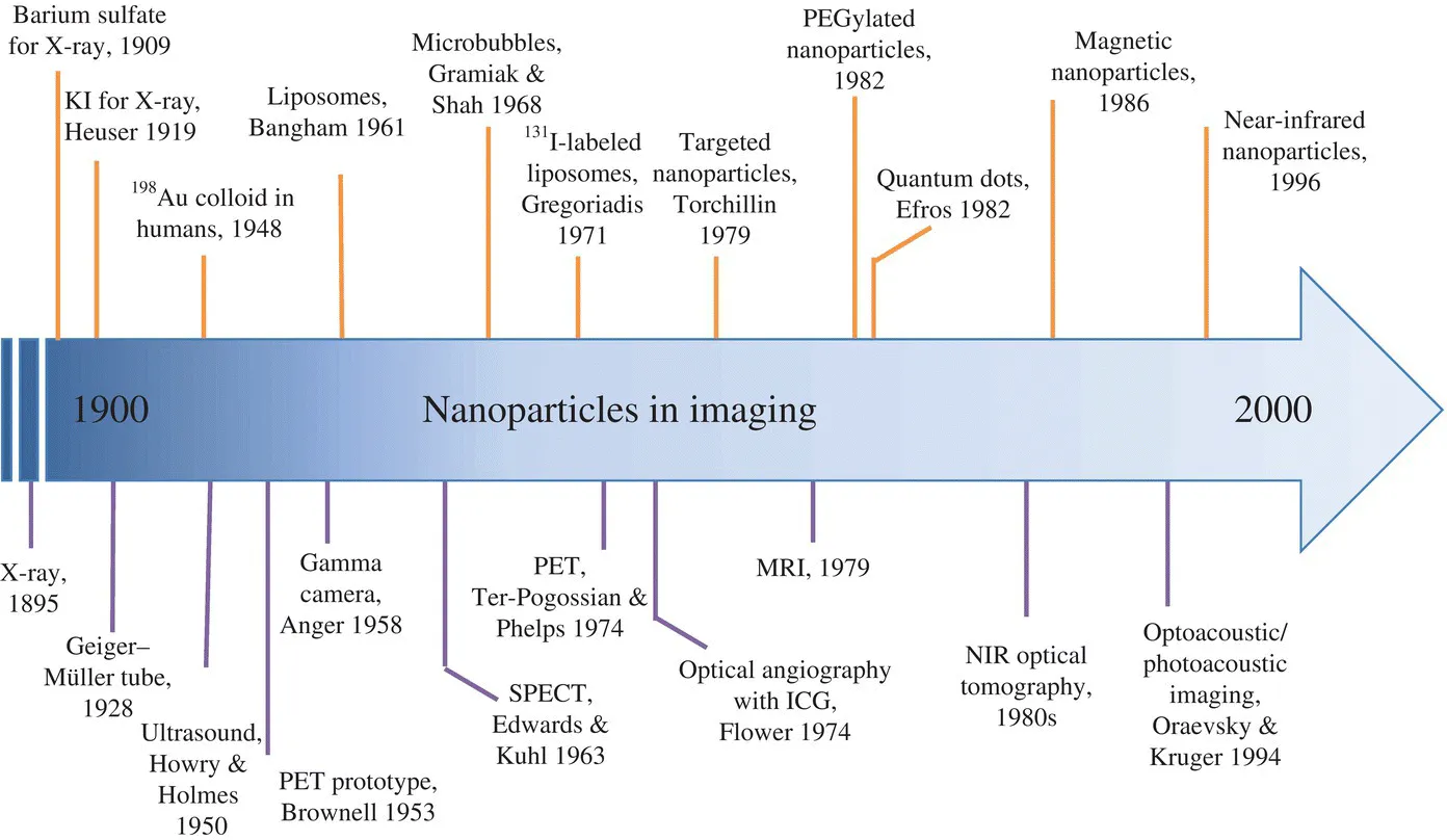
From the onset of radiology and the first contrast agents to the end of the twentieth century, imaging techniques such as X-ray, PET, SPECT, ultrasound, MRI, optical, and photoacoustics have emerged. The first imaging nanoparticles appeared only in the middle of the twentieth century. The progress and the application of imaging nanoparticles followed the advent of new imaging modalities and diverged into two equally important directions. In one direction, de novo nanoparticle designs were developed for specific imaging modalities. Some examples include magnetic particles for MRI, quantum dots (QDs) for optical, and nanobubbles for ultrasound. The other direction adopted previously established designs of nanoparticles (for instance, for drug delivery) and modified them for imaging applications. Some examples include liposomes, virions, cross-linked nanoparticles, and surface modification to increase the nanoparticles' imaging specificity. Regardless of direction, many nanoparticles applications often began as unexpected discoveries. Many steps to refine their design were necessary to turn them from a mere curiosity to a clinically acceptable tool. Today, the continued improvement in nanoparticle synthesis, conjugation technique, and novel biomarkers made the nanoparticle approach a unique and well-differentiated scientific direction that blends seamlessly with clinical imaging. The historical trend illustrated in Figure 1.2 highlights the most important milestones toward this direction and is discussed in this chapter.
Figure 1.2 Timeline of the most important events in the development of nanoparticles for imaging and diagnostics covering the period from the twentieth century. The upper part corresponds to nanoparticles, and the lower part to the development of imaging modalities.
1.2 X-Ray and First Contrast Agents (1895–1930s)
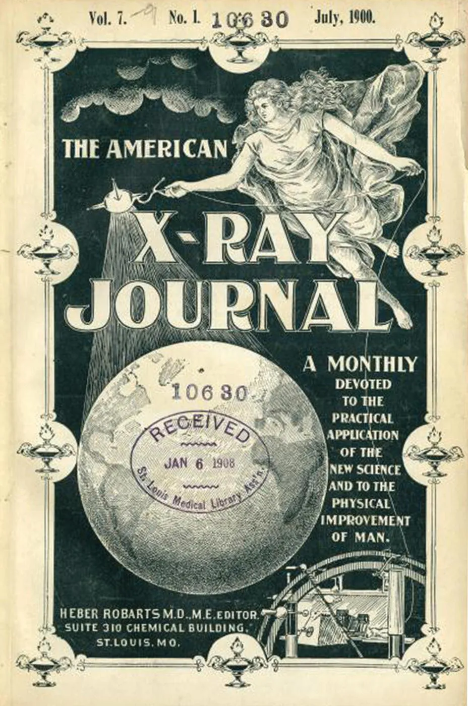
The history of medical imaging started on November 8, 1895, when a 50-year-old Wilhelm Conrad Röntgen—a physicist from the University of Würzburg in Germany—observed a greenish glow from a recently invented Crookes tube. A new form of radiation, which Röntgen called an “X-ray,” freely penetrated through biological tissue but was absorbed by dense material such as bones. Recorded on radiation-sensitive photographic plates, a well-recognized X-ray image was made. This entirely noninvasive imaging technique quickly spread across the world after its demonstration to the public in 1896. A review of major medical colleges across the United States conducted by the American X-Ray Journal (Fig. 1.3 shows the cover of this journal) in 1899 revealed more than 80 institutions where X-ray machines were available for patients [1], a remarkable rate given that it was just 4 years after X-ray discovery. With X-ray imaging, bone fractures, kidney stones, and metallic objects such as bullets and needles could be reliably located. With further refinement, physicians could even recognize and visualize certain organs. However, imaging inside the organs was impossible since the low and uniform density of soft tissue composed of transparent to X-rays water and organic media provided little contrast within the tissue.
Figure 1.3 The American X-Ray Journal established in May 1897 was one of the first imaging journals. Launched by Dr. H. Robarts, a prominent radiologist from St. Louis, his biography is described in Ref. [2]. The journal existed until 1905.
(Courtesy of Becker Library, Washington University School of Medicine.)
To address this shortcoming, W. Cannon from Harvard Medical School began developing “contrast agents,” biocompatible compounds that could absorb X-rays. In 1905, he discovered that high-density metal salts such as bismuth-based compounds provided the desired contrast in the intestines: “The animals thus fed with food mixed with bismuth subnitrate were exposed to the X-rays and, without disturbing the processes of digestion, the movements of the food in the stomach and small intestine were observed by means of the shadows cast on a fluorescent screen” [3]. A few years later, a less toxic barium sulfate mixed with foodstuffs became the first broadly used contrast agent in X-ray imaging of the digestive tract [4]. This water-insoluble salt (to prevent barium toxicity) was swallowed with food prior to the imaging procedure to outline the esophagus, stomach, and small intestines. The contrast could also be inserted via enemas to visualize the colon. This practice allowed the visualization of tumors, strictures, blockages, and ulcers and has been so simple and successful that it is still in use today.
The next advancement in the development of contrast agents came from Argentina, where in 1919 the radiologist Dr. C. Heuser intravenously injected a water-soluble potassium iodide to image the circulatory system. High-density iodide provided significant attenuation to X-ray radiation, causing the blood vessels to appear lighter on film. A few years later, Heuser utilized another iodinated compound called Lipiodol synthesized in 1901 by the French chemist M. Guerbet. Lipiodol is a low-viscosity radio-opaque diagnostic agent formed by the iodination of the fatty acids in poppy-seed oil and was applied to investigate the uterine cavity and fallopian tubes. Due to its high density and low toxicity, many iodinated compounds are commonly used today in X-ray and computer tomography (CT) imaging—a successor of the X-ray technique. (One of the leading companies of X-ray contrast agents is the Guerbet Group established by the son of Lipiodol's inventor in 1926.) However, despite several decades of continuous efforts to improve X-ray instrumentation and expand X-ray imaging to soft tissue with contrast agents, diagnosing diseases of internal organs suffered from unacceptably low contrast. New technologies were desperately needed.
1.3 Rise of the Nuclear Imaging Techniques (1940s–1950s)
Shortly after World War II in 1946, the U.S. Congress passed the Atomic Energy Act that transferred nuclear weapon development and nuclear power management to civilian, rather than military control. The Oak Ridge Laboratory in Tennessee was directed to provide radioisotopes for peaceful purposes, especially for medical applications. One of the first isotopes made available was 198Au colloid. It was produced by bombarding gold foil w...
Table of contents
Cover
Title page
Copyright page
Dedication page
Contributors
Preface
Acknowledgments
1 Historical Perspective on Nanoparticles in Imaging from 1895 to 2000
Part I: Nanoparticle Design, Synthesis and Characterization
Part II: Imaging Modalities: from Concepts to Applications
Part III: Nanotechnology in Biomedical Imaging and Beyond
Index
End User License Agreement
Frequently asked questions
Yes, you can cancel anytime from the Subscription tab in your account settings on the Perlego website. Your subscription will stay active until the end of your current billing period. Learn how to cancel your subscription
No, books cannot be downloaded as external files, such as PDFs, for use outside of Perlego. However, you can download books within the Perlego app for offline reading on mobile or tablet. Learn how to download books offline
Perlego offers two plans: Essential and Complete
Essential is ideal for learners and professionals who enjoy exploring a wide range of subjects. Access the Essential Library with 800,000+ trusted titles and best-sellers across business, personal growth, and the humanities. Includes unlimited reading time and Standard Read Aloud voice.
Complete: Perfect for advanced learners and researchers needing full, unrestricted access. Unlock 1.5M+ books across hundreds of subjects, including academic and specialized titles. The Complete Plan also includes advanced features like Premium Read Aloud and Research Assistant.
Both plans are available with monthly, semester, or annual billing cycles.
We are an online textbook subscription service, where you can get access to an entire online library for less than the price of a single book per month. With over 1.5 million books across 990+ topics, we’ve got you covered! Learn about our mission
Look out for the read-aloud symbol on your next book to see if you can listen to it. The read-aloud tool reads text aloud for you, highlighting the text as it is being read. You can pause it, speed it up and slow it down. Learn more about Read Aloud
Yes! You can use the Perlego app on both iOS and Android devices to read anytime, anywhere — even offline. Perfect for commutes or when you’re on the go. Please note we cannot support devices running on iOS 13 and Android 7 or earlier. Learn more about using the app
Yes, you can access Nanotechnology for Biomedical Imaging and Diagnostics by Mikhail Y. Berezin in PDF and/or ePUB format, as well as other popular books in Technology & Engineering & Applied Sciences. We have over 1.5 million books available in our catalogue for you to explore.