Instant Anatomy presents anatomy and anatomical relationships in a simple, unique, schematic manner to aid the speedy understanding and retrieval of anatomical facts. It shows structures such as nerves and blood vessels in their entirety, unlike the partial, regional presentations given in most textbooks.
Covering the major aspects of anatomy, each section presents the relevant structures in double page spreads, with clear, full-colour diagrams on the left and concise text for each structure on the right. This new fifth edition includes more surface anatomy such as new myotome maps, bones of the hands and feet, principles of movement at shoulder and hip and images to clarify the understanding of the inguinal region and the lesser sac of the stomach.
Ideal for use alongside a core anatomy textbook, Instant Anatomy is the perfect quick reference guide for medical students, surgeons, radiologists and those in many other specialties. The companion website at www.instantanatomy.net with its podcasts and wide ranging multiple choice questions provide invaluable exam preparation.
- English
- ePUB (mobile friendly)
- Available on iOS & Android
eBook - ePub
Instant Anatomy
About this book
Trusted by 375,005 students
Access to over 1 million titles for a fair monthly price.
Study more efficiently using our study tools.
Information
1: Arteries
- Coronary arteries
- Ascending & arch of aorta
- Internal carotid artery, vertebrobasilar system & circle of Willis
- Ophthalmic artery
- External carotid artery
- Maxillary artery
- Middle meningeal artery
- Subclavian artery
- Axillary artery
- Brachial artery
- Radial artery
- Ulnar artery
- Thoracic (descending) aorta
- Abdominal aorta
- External iliac artery
- Coeliac trunk
- Superior mesenteric artery
- Inferior mesenteric artery
- Internal iliac artery
- Femoral artery
- Popliteal artery
- Anterior tibial artery
- Posterior tibial artery
- Fibular (peroneal) artery
- Arterial anastomoses around scapula
- Arterial anastomoses around hip
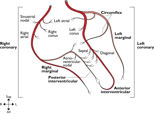
Coronary arteries
Coronary Arteries
From: Ascending aorta
To: Myocardium
Right coronary artery. Originates from the anterior aortic sinus. It passes anteriorly between the pulmonary trunk and the right auricle to reach the atrioventricular sulcus in which it runs down the anterior surface of the right cardiac border and then onto the inferior surface of the heart. It terminates at the junction of the atrioventricular sulcus and the posterior interventricular groove by anastomosing with the circumflex branch of the left coronary artery and giving off the posterior interventricular (posterior descending) artery. It supplies the right atrium and part of the left atrium, the sinuatrial node in 60% of cases, the right ventricle, the posterior part of the interventricular septum and the atrioventricular node in 80% of cases.
Left coronary artery. Arises from the left posterior aortic sinus. It passes laterally, posterior to the pulmonary trunk and anterior to the left auricle to reach the atrioventricular groove where it divides into an anterior interventricular (formally left anterior descending) artery and circumflex branches. The circumflex artery runs in the atrioventricular sulcus around the left border of the heart to anastomose with the right coronary artery. The anterior interventricular artery descends on the anterior surface of the heart in the anterior interventricular groove and around the apex of the heart into the posterior interventricular groove where it anastomoses with the posterior interventricular branch of the right coronary artery. The left coronary artery supplies the left atrium, left ventricle, anterior interventricular septum, sinuatrial node in 40% of cases and the atrioventricular node in 20%.
Dominance. In approximately 10% of hearts the posterior interventricular artery arises from the circumflex artery (left coronary) and then most of the left ventricle and interventricular septum are supplied by the left coronary artery. The heart is said to have left cardiac dominance.
Ascending & arch of aorta
Ascending & Arch of Aorta
From: Left ventricle
To: Descending aorta
Ascending aorta. Arises at the vestibule of the left ventricle at the level of the third left costal cartilage and passes upwards and slightly to the right to a point behind the sternum at the level of the manubriosternal joint (second costal cartilage) where it becomes the arch of the aorta. It is enclosed in fibrous and serous pericardium. Anterior to it are the right auricle, the infundibulum of the right ventricle and pulmonary trunk. Posterior, lie the left atrium, the right pulmonary artery and right main bronchus. To the left lie the pulmonary trunk and the left auricle. To the right are the superior vena cava and the right atrium.
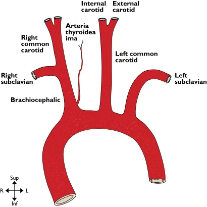
Arch of aorta. The arch begins posterior to the manubriosternal joint at the level of the second costal cartilage and passes posterior and to the left, over the left main bronchus to end at the left side of the body of T4 vertebra. Its highest level is the mid-point of the manubrium sterni and at this level its three main branches emerge. Anterior and to the left of the arch are (from anterior to posterior) the left phrenic nerve, vagal and sympathetic contributions to the cardiac plexus, and the left vagus. Also, the left superior intercostal vein runs forwards on the arch anterior to the vagus and posterior to the phrenic nerve. Lateral to all these structures are the pleura and left lung. Posterior and to the right of the arch are the trachea, deep cardiac plexus, left recurrent laryngeal nerve, oesophagus, thoracic duct and the body of T4. Inferior to the arch are the pulmonary bifurcation, the left main bronchus, the ligamentum arteriosum and the left recurrent laryngeal nerve. From its superior surface emerge the brachiocephalic artery, the left common carotid and left subclavian arteries. Within the adventitia of the ascending and arch of the aorta lie baro- and chemoreceptors.
Brachiocephalic artery. Arises from the convexity of the aortic arch behind the manubrium sterni and passes upwards and posteriorly to the right. It divides into the right subclavian and right common carotid arteries posterior to the right sternoclavicular joint. Anterior to it are the left brachiocephalic vein with the right inferior thyroid vein entering it, and the thymic remnants. The artery initially lies anterior to the trachea and then passes to lie on its right lateral side. On the right of the artery are the right brachiocephalic vein, upper part of the superior vena cava, the pleura and the cardiac branches of the vagus. The main vagal trunk is more posterolateral. At the origin of the brachiocephalic artery the left common carotid artery lies posteriorly on its left.
Ascending & arch of aorta
Common carotid arteries. The right common carotid artery arises from the brachiocephalic artery as it divides posterior to the right sternoclavicular joint, whilst the left common carotid arises from the convexity of the aortic arch. Both end as the arteries bifurcate at the level of the upper border of the thyroid cartilage (C4).
Left common carotid artery (thorax). Lying anterior to the thoracic part of this artery are the left brachiocephalic vein and the thymic remnant. Posterior to it in its lower part are the left subclavian artery and the trachea whilst further superiorly there is the left recurrent laryngeal nerve, the thoracic duct and the left side of the oesophagus. On its right at its origin is the brachioceph...
Table of contents
- Cover
- The Authors
- Title Page
- Copyright
- Preface to Fifth Edition
- Preface to First Edition
- Notes on the Text
- Chapter 1: Arteries
- Chapter 2: Veins
- Chapter 3: Lymphatics
- Chapter 4: Autonomic Nervous System
- Chapter 5: Cranial Nerves
- Chapter 6: Peripheral Nerves
- Chapter 7: Dermatomes and Cutaneous Nerve Distribution
- Chapter 8: Muscles
- Chapter 9: Joints
- Chapter 10: Ossification Times
- Chapter 11: Foramina—Skull and Spine
- Chapter 12: Position of Structures According to Vertebral Levels Position of Structures According to Vertebral Levels
- Chapter 13: Pharyngeal Derivatives
- Chapter 14: Surface Anatomy and Key Areas
- End User License Agreement
Frequently asked questions
Yes, you can cancel anytime from the Subscription tab in your account settings on the Perlego website. Your subscription will stay active until the end of your current billing period. Learn how to cancel your subscription
No, books cannot be downloaded as external files, such as PDFs, for use outside of Perlego. However, you can download books within the Perlego app for offline reading on mobile or tablet. Learn how to download books offline
Perlego offers two plans: Essential and Complete
- Essential is ideal for learners and professionals who enjoy exploring a wide range of subjects. Access the Essential Library with 800,000+ trusted titles and best-sellers across business, personal growth, and the humanities. Includes unlimited reading time and Standard Read Aloud voice.
- Complete: Perfect for advanced learners and researchers needing full, unrestricted access. Unlock 1.4M+ books across hundreds of subjects, including academic and specialized titles. The Complete Plan also includes advanced features like Premium Read Aloud and Research Assistant.
We are an online textbook subscription service, where you can get access to an entire online library for less than the price of a single book per month. With over 1 million books across 990+ topics, we’ve got you covered! Learn about our mission
Look out for the read-aloud symbol on your next book to see if you can listen to it. The read-aloud tool reads text aloud for you, highlighting the text as it is being read. You can pause it, speed it up and slow it down. Learn more about Read Aloud
Yes! You can use the Perlego app on both iOS and Android devices to read anytime, anywhere — even offline. Perfect for commutes or when you’re on the go.
Please note we cannot support devices running on iOS 13 and Android 7 or earlier. Learn more about using the app
Please note we cannot support devices running on iOS 13 and Android 7 or earlier. Learn more about using the app
Yes, you can access Instant Anatomy by Robert H. Whitaker,Neil R. Borley in PDF and/or ePUB format, as well as other popular books in Medicine & Anatomy. We have over one million books available in our catalogue for you to explore.