![]()
Part 1
The ear, hearing and balance
![]()
1
The ear: applied basic science
Divisions of the ear
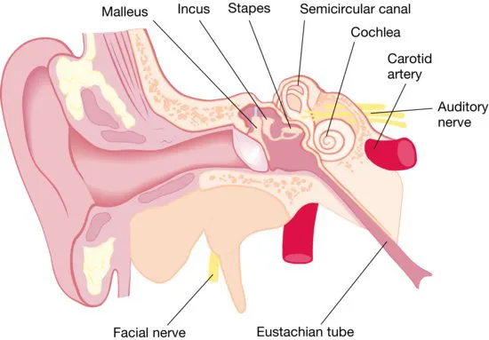
The ear is usually described as comprising three parts – the
external (outer),
middle and
inner ear (
Fig. 1.1). The external ear is made up of the pinna and the external ear canal or ‘external auditory meatus'.
The pinna
The pinna (auricle) is composed of cartilage. This is covered with closely adherent perichondrium which gives it its blood supply and with skin. The head and neck in the embryo develops from a number of primitive tissue units known as the pharyngeal arches and the pinna is derived from the fusion of six tubercles of the first of these arches. This is a complex process and anomalies such as fistulas, accessory auricles and deformity of the ear can result from failure of fusion of these tubercles.
The external auditory meatus or ear canal
The external auditory meatus is about 25 mm in length. It has a skeleton of cartilage in its outer third (where it contains hairs and ceruminous or ‘wax-producing' glands) and bone in its inner two-thirds. The skin of the inner part is thin, adherent and sensitive. Wax, debris or foreign bodies may easily lodge at the medial end of the meatus.
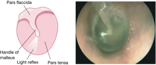
The tympanic membrane or eardrum (Fig. 1.2)
The tympanic membrane is composed of three layers from out to in – skin, fibrous tissue and mucosa. The normal appearance of the membrane is pearly and opaque. When light reflects off the drum it forms a characteristic triangular ‘light reflex' due to its concave shape. If you see this ‘light reflex' that is good evidence that the drum is normal.
The tympanic cavity or middle ear
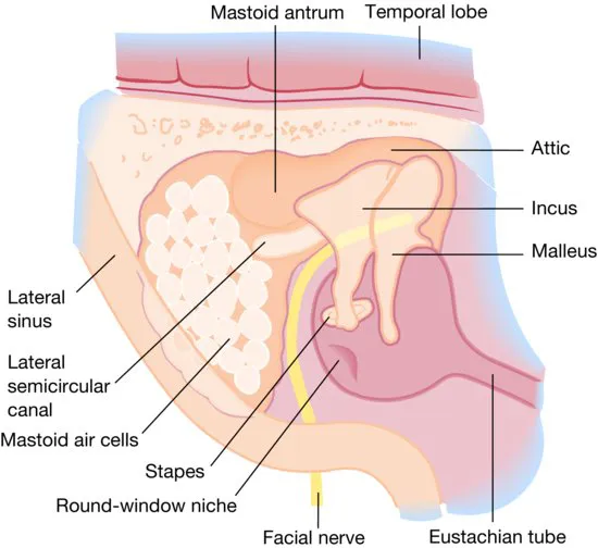
Medial to the eardrum, the tympanic cavity is an air-containing space 15 mm high and 15 mm antero-posteriorly, although only 2 mm deep in parts. The middle ear contains the small middle-ear bones or ossicles- the malleus, incus and stapes (‘hammer', ‘anvil' and ‘stirrup') (Figs 1.1 and 1.3). Its medial wall is crowded with structures closely related to one another: the facial nerve, the round and oval windows, the lateral semicircular canal and the cochlea.
The Eustachian tube
The Eustachian tube connects the middle ear with the nasopharynx at the back of the nasal cavity. The tube permits aeration of the middle ear and if it is obstructed fluid may accumulate in the middle ear causing deafness. The tube is shorter, wider and more horizontal in the infant than in the adult. Secretions or food may enter the tympanic cavity more easily when the baby is supine particularly during feeding. The tube is normally closed and opens on swallowing because of movement of the muscles of the palate. This movement is impaired in cleft palate children who often develop accumulation of middle-ear fluid (otitis media with effusion).
The inner ear
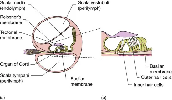
The inner ear is made up of the cochlea, responsible for hearing and the semi-circular canals which house the ‘balance organs'. The delicate neuroepithelium is well protected in the temporal bone of the skull (Fig. 1.4).
The facial nerve
The facial nerve is the motor nerve to the muscles of facial expression. Intimately associated with the ear, it is embedded in the temporal bone and passes through the middle ear but exits the skull at the stylomastoid foramen just in front of the mastoid process (Fig. 1.3). In infants, the mastoid process is undeveloped and the nerve is very superficial.
The mastoid cells
The mastoid cells form a honeycomb within the temporal bone, acting as a reservoir of air to limit pressure changes within the middle ear. The extent of pneumatization is very variable and is usually reduced in chronic middle ear disease when the mastoid is often said to be ‘sclerotic'.
The mechanism of hearing (Fig. 1.4)
Sound causes the eardrum to vibrate. This energy is transmitted via the ossicles to the oval window which is in contact with the stapes. A ‘travelling wave' is set up in the fluids of the inner ear. Specialized neuroepithelial cells (‘hair cells') in the cochlea or inner ear convert this energy to nerve impulses which then travel along the auditory pathway to the cortex where they are recognized as sound. Diseases which interfere with transmission of sound across the outer and middle ear cause conductive deafness, and diseases in the inner ear which interfere with the conversion of this energy to nerve impulses or with the transmission of these nerve impulses cause sensorineural or ‘nerve' deafness.
- The facial nerve is intimately related to the middle and inner ears. Always check the ear carefully in a patient with facial palsy.
- The middle ear amplifies sound. The inner ear is essential for hearing. Middle-ear disease may cause some degree of deafness but if the inner ear is not functioning the patient will be completely deaf in that ear.
Go to
www.lecturenoteseries.com/ENT to test yourself using the interactive MCQs.
![]()
2
Clinical examination of the ear
The examination of the ...