Mastery of ECG interpretation is achieved not only by pattern recognition, but equally importantly, by a clear, practical understanding of how electricity moves through the heart and how disruption of that movement manifests itself via ECG tracings.
ECGs for Beginners, written by one of the world's most respected electrophysiologists with over 40 years experience of training clinicians, will provide cardiology and electrophysiology trainees with an easy to follow, step-by-step guide to the topic, thus enabling them to both understand and interpret ECG readings in order to to best manage their patients.
Packed with over 250 high-quality ECG tracings, as well as management algorithms and key points throughout, every chapter also contains self-assessment questions, allowing the reader to test themselves on what they've just learnt.
All kinds of arrhythmias will be covered, as well as morphological abnormalities such as atrial and ventricular problems. Importantly, normal ECG readings will be presented alongside abnormal readings, to best demonstrate how and why abnormalities occur.
ECGs for Beginners is an essential purchase for all cardiology and electrophysiology trainees, as well as being a handy refresher guide for the experienced physician.
- English
- ePUB (mobile friendly)
- Available on iOS & Android
eBook - ePub
ECGs for Beginners
About this book
Trusted by 375,005 students
Access to over 1 million titles for a fair monthly price.
Study more efficiently using our study tools.
Information
PART I
The Normal Electrocardiogram
In the first chapter the anatomical and electrophysiological bases essential to understanding the human electrocardiogram (ECG), are outlined. Chapter 2 explains how the ECG records the path of cardiac activation through the heart from the sinus node to the ventricular muscle in the form of activation curves (depolarization and repolarization) of the atria (P waves) and ventricles (the QRS-T complex). Chapter 3 describes ECG devices and recording techniques. Lastly, Chapter 4 explains in detail the process for interpreting normal and pathologic ECG recordings, including the normal characteristics of each parameter studied.
A full understanding of these concepts is essential before continuing on to the other parts of the book. Please start the first four chapters again if necessary.
CHAPTER 1
Anatomical and Electrophysiological Bases
1.1. The Heart Walls
The heart has four cavities, two atria and two ventricles, comprised mainly of contractile cells called cardiomyocytes. The electrical stimulus originating in the sinus node (SN) is distributed through the entire heart by means of a specific conduction system (SCS).
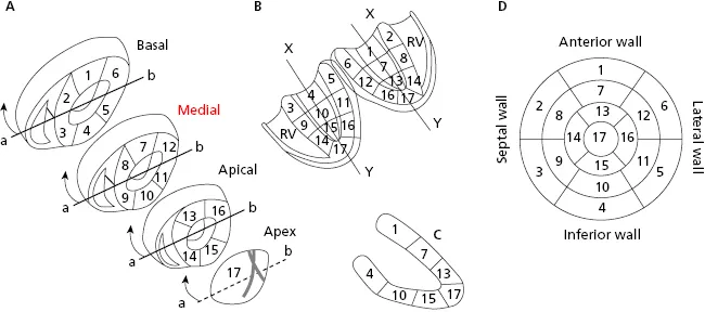
The left ventricle (LV) has four walls: anterior, septal, inferior, and lateral. Figure 1.1 shows the three segments of the anterior and inferior walls, the five segments of the septal and lateral walls, and the apex segment (segment 17). Magnetic resonance imaging has now shown that the previously-named posterior wall corresponds to the inferobasal segment of the inferior wall (segment 4 in Fig. 1.1) (Bayés de Luna et al., 2006a; Bayés de Luna A and Fiol-Sala, 2008). [A]
1.2. Coronary Circulation (Fig. 1.2)
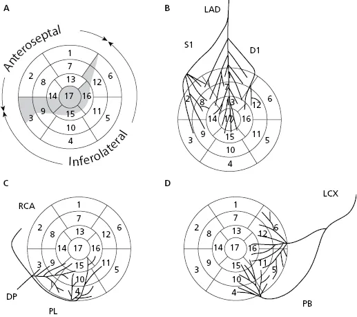
Based on coronary perfusion, the heart is divided into two zones: the anteroseptal zone, perfused by the left anterior descending artery (LAD) (Fig. 1.2A) and the inferolateral zone, perfused by the right coronary artery (RCA) and circumflex artery (CX) (Figs 1.2C and 1.2D). The heart has areas of shared perfusion (shown in grey in Fig. 1.2A) in which one of the two arteries dominates. For example, segment 17 (apex) is perfused by the LAD, if long; otherwise by the RCA and even partially by the CX. [B]
1.3. The Specific Conduction System (Fig. 1.3)
Electrical stimuli pass through the internodal pathways (Bachmann, Wenckebach and Thorel bundles), from the sinus node to the AV node and the His bundle. From there stimuli reach the ventricles through the ventricular conduction system: the right branch (RB) and the trunk of the left branch (LB), and its divisions (superoanterior and inferoposterior fascicles and the middle fibers that exist between them) (Figs 1.3A and 1.3B). [C]
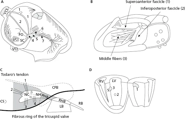
Table of contents
- Cover
- Table of Contents
- Title page
- Copyright page
- Preface
- Forewords to Previous Editions
- Foreword
- PART I: The Normal Electrocardiogram
- PART II: Morphological Abnormalities in the ECG
- PART III: The ECG in Arrhythmias
- PART IV: ECG in Clinical Practice
- Bibliography
- Supplemental Images
- Index
- End User License Agreement
Frequently asked questions
Yes, you can cancel anytime from the Subscription tab in your account settings on the Perlego website. Your subscription will stay active until the end of your current billing period. Learn how to cancel your subscription
No, books cannot be downloaded as external files, such as PDFs, for use outside of Perlego. However, you can download books within the Perlego app for offline reading on mobile or tablet. Learn how to download books offline
Perlego offers two plans: Essential and Complete
- Essential is ideal for learners and professionals who enjoy exploring a wide range of subjects. Access the Essential Library with 800,000+ trusted titles and best-sellers across business, personal growth, and the humanities. Includes unlimited reading time and Standard Read Aloud voice.
- Complete: Perfect for advanced learners and researchers needing full, unrestricted access. Unlock 1.4M+ books across hundreds of subjects, including academic and specialized titles. The Complete Plan also includes advanced features like Premium Read Aloud and Research Assistant.
We are an online textbook subscription service, where you can get access to an entire online library for less than the price of a single book per month. With over 1 million books across 990+ topics, we’ve got you covered! Learn about our mission
Look out for the read-aloud symbol on your next book to see if you can listen to it. The read-aloud tool reads text aloud for you, highlighting the text as it is being read. You can pause it, speed it up and slow it down. Learn more about Read Aloud
Yes! You can use the Perlego app on both iOS and Android devices to read anytime, anywhere — even offline. Perfect for commutes or when you’re on the go.
Please note we cannot support devices running on iOS 13 and Android 7 or earlier. Learn more about using the app
Please note we cannot support devices running on iOS 13 and Android 7 or earlier. Learn more about using the app
Yes, you can access ECGs for Beginners by Antoni Bayés de Luna in PDF and/or ePUB format, as well as other popular books in Medicine & Physiology. We have over one million books available in our catalogue for you to explore.