Discussing recent findings, up-to-date research, and novel strategies, the book integrates perspectives from pharmacology, toxicology, and biochemistry to illustrate the potential of lysosomes in drug discovery and development.
• Explores basic principles and properties of lysosomes that allow them to act as regulators of cell metabolism, therapeutic targets, and sites for activation of drug conjugates
• Discusses the role of lysosomes in metabolism, drug targeting, apoptosis, cancer, aging, inflammation, autophagy, metabolism, toxicity, and membrane repair
• Introduces new pathways in therapeutic development and new mechanisms in drug development
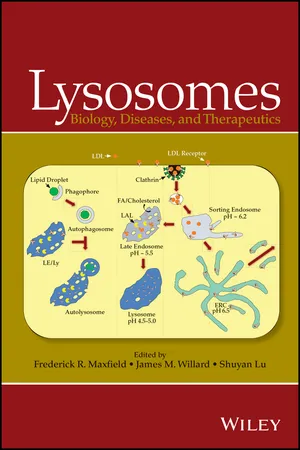
eBook - ePub
Lysosomes
Biology, Diseases, and Therapeutics
- English
- ePUB (mobile friendly)
- Available on iOS & Android
eBook - ePub
Lysosomes
Biology, Diseases, and Therapeutics
About this book
Information
Chapter 1
Lysosomes: An Introduction
Frederick R. Maxfield
Department of Biochemistry, Weill Cornell Medical College, New York, NY, USA
Late endosomes and lysosomes (LE/Ly) are the main digestive organelles of eukaryotic cells. They contain a variety of enzymes and accessory proteins [1] that are capable of the hydrolysis of many biological molecules. The LE/Ly are maintained at an acidic pH, and most lysosomal enzymes are acid hydrolases with acidic pH optima. Substrates are delivered to LE/Ly either by endocytic processes (e.g., receptor-mediated endocytosis, pinocytosis, and phagocytosis) or by autophagic processes in which autophagosomes containing cellular content fuse with the LE/Ly.
The history of lysosomal studies has been discussed in several publications [2–5]. The study of lysosomal digestion began in the late 19th century with microscopic observations of cells ingesting material. In the same period, physicians began to see abnormal storage of material in pathology specimens from patients with unusual illnesses. In the 1950s and 1960s, lysosomes were purified by differential centrifugation techniques, and the linkage was established between these organelles and the microscopic observations of storage material in what then became known as lysosomal storage diseases. These discoveries were followed by contributions from many laboratories that have led to a fairly detailed understanding of the biogenesis, composition, and function of lysosomal organelles.
There have been several recent advances in our understanding of lysosomal function. In particular, we now have a much better understanding of the mechanisms underlying the regulation of lysosome formation (see Chapters 2 and 7). In addition, while autophagy has been known for many years, there have been dramatic increases in our understanding of its molecular mechanisms in the past decade (see Chapter 2).
A brief note about nomenclature may be useful. As with other organelles, the acidic digestive organelles are heterogeneous and undergo rapid changes due to various membrane trafficking processes. Several years ago, a definition was proposed [6] in which the organelles that were actively receiving new lysosomal enzymes from the trans-Golgi network would be called late endosomes, while more mature acidic digestive organelles would retain the name lysosomes. This distinction remains useful in describing two broad groups of acidic digestive organelles, but it does lead to some semantic difficulties. For example, in many cells, digestion of endocytosed proteins by lysosomal hydrolases occurs mainly in the late endosomes. Thus, “lysosomal digestion” is nearly complete before the endocytosed material can enter “lysosomes.” In addition, even within the same cell there can be significant differences in the properties (size, morphology, enzyme content, substrate content, etc.) in organelles that are broadly grouped as late endosomes or lysosomes. These difficulties arise because of the intrinsically dynamic nature of these organelles, which are rapidly exchanging membrane and lumenal content with each other and with other organelles. Unfortunately, a completely accurate and descriptive terminology may not be possible.
1.1 Historical Background
The study of lysosomes began with the microscopic observations summarized by Metchnikoff [7] in the late 19th century. He and his contemporaries reported their direct microscopic analysis on the uptake of foreign particles into cells, and the subsequent digestion of these particles. In some cases, Metchnikoff observed that ingested pieces of litmus would change from blue to red, implying an acid environment surrounding the ingested particles. Metchnikoff emphasized that aspects of phagocytic uptake were evolutionarily conserved from single cell organisms through leukocytes obtained from mammals.
One of the ongoing themes in studies of lysosomes is that there is a dynamic interplay between the study of diseases and the contributions of basic science. In parallel with the developments in the cell biology of digestion of internalized material, there were descriptions beginning in the 19th century of various storage diseases in which cellular accumulations of material were observed upon pathological examination of tissues from patients with a variety of diseases [3]. These included Gaucher, Tay–Sachs [8], and Niemann–Pick diseases among others. Many of these storage disorders were recognized to have a hereditary component, and several of them were also recognized to lead to accumulation of certain molecular species, especially lipids.
The modern era of lysosome study began with the purification of lysosomes by Christian de Duve and his colleagues in the 1950s, and a review of these discoveries presents many of the important details [5]. De Duve's laboratory sought to understand the localization of glucose-6-phosphatase in liver fractions as part of study of the mechanisms of insulin action. Among the phosphatase activities that they studied was acid phosphatase, and they noticed that its activity was latent and could only be observed when membranes were disrupted. At first they considered that this might suggest a mitochondrial localization, but improved centrifugation techniques led to separation of the acid phosphatase fraction from the mitochondria. In 1955, they published a paper describing isolation of particles that contained five enzyme activities with acidic pH optima [9]. Alex Novikoff had also been working on the distribution of various enzymes in cells. After visiting de Duve's laboratory, Novikoff carried out the first electron microscopic studies of the newly isolated organelles. To do this, he would carry fresh samples from the de Duve laboratory in Belgium to the new electron microscope available in the laboratory of Albert Claude in Paris [10]. He observed that the fractions were enriched in dense bodies about 370 nm in length, and he also noted that they resembled in many ways similar structures observed in hepatocytes [11]. Working independently, Straus [12] isolated “droplets” from rat kidneys and showed that these were enriched in acid phosphatase and other enzymes [13]. Straus also showed that similar, but larger, droplets were observed after intraperitoneal injection of egg white and reported that the injected protein could be found in these droplets. This linked the degradative organelles with the uptake of extracellular material.
After the discovery of lysosomes by de Duve and his coworkers, the idea that lysosomal defects might be the underlying cause of the storage diseases began to be considered. The clear demonstration of association of these storage disorders with lysosomes was first made by Hers [14, 15] when the defect in a glycogen storage disease was shown to be a deficiency in the lysosomal enzyme acid maltase. This was followed by the association of several of the storage disorders, now classified as lysosomal storage disorders, with a deficiency in specific enzymes.
Work from Neufeld and others [16] showed that enzymes secreted by one cell could be added to the culture medium of a cell lacking a particular enzyme and correct the storage defect. It was found, however, that enzymes secreted from I-cell disease fibroblasts could not correct the enzyme deficiencies in other cells. This and other observations led to the hypothesis that there must be a tag of some type on lysosomal enzymes and there must be receptors on the surface of cells that could selectively endocytose the tagged enzymes [4]. Shortly thereafter, it was found that the uptake of β-glucuronidase could be inhibited by mannose-6-phosphate and that the uptake of the enzyme could also be blocked by pretreatment with a phosphatase [17]. Mannose-6-phosphate glycoconjugates were then identified on many lysosomal enzymes.
The cation-independent mannose-6-phosphate receptor was first isolated from bovine liver in 1981 [18], and the sequence of the human receptor was determined in 1988 [19]. It was found to be identical to an independently identified receptor for insulin-like growth factor II (IGF-II) [20]. A second mannose-6-phosphate receptor (the cation-dependent mannose-6-phosphate receptor) was identified in 1985 [21].
In 1978, Ohkuma and Poole [22], who were colleagues of de Duve, used the uptake of fluorescein–dextran into lysosomes to measure the pH of these organelles accurately based on the pH dependence of fluorescein fluorescence. They also showed that lysosomal acidification required ATP and that weak bases could increase the pH of these organelles. Most of the lysosomal hydrolases have acidic pH optima, and it seems likely that this pH dependence is protective for events that lead to disruption of lysosomes since the enzymes will have greatly reduced activity at cytoplasmic or extracellular pH.
From the 1980s to the present, there has been an explosive growth in the number of studies of various aspects of lysosome biology, and many aspects of lysosomal biogenesis and function are becoming well understood. It is particularly gratifying that understanding of the basic biochemistry and cell biology is leading to therapies for several of the lysosomal storage disorders. In addition, understanding of endocytic targeting to digestive organelles is leading to development of very selectively targeted delivery of therapeutic agents. Many of these studies are summarized in the chapters of this book.
References
- [1] Lubke T, Lobel P, Sleat DE. Proteomics of the lysosome. Biochim Biophys Acta 2009;1793:625–635.
- [2] Holtzman E. Lysosomes. New York: Plenum; 1989. 439 p.
- [3] Platt FM, Walkley SU. Lysosomal Disorders of Brain: Recent Advances in Molecular and Cellular Pathogenesis and Treatment. Oxford; New York: Oxford University Press; 2004. xxvii, 44...
Table of contents
- Cover
- Title Page
- Copyright
- Table of Contents
- Preface
- List of Contributors
- Chapter 1: Lysosomes: An Introduction
- Chapter 2: Lysosome Biogenesis and Autophagy
- Chapter 3: Multivesicular Bodies: Roles in Intracellular and Intercellular Signaling
- Chapter 4: Lysosomes and Mitophagy
- Chapter 5: Lysosome Exocytosis and Membrane Repair
- Chapter 6: Role of Lysosomes in Lipid Metabolism
- Chapter 7: TFEB, Master Regulator of Cellular Clearance
- Chapter 8: Lysosomal Membrane Permeabilization in Cell Death
- Chapter 9: The Lysosome in Aging-Related Neurodegenerative Diseases
- Chapter 10: Lysosome and Cancer
- Chapter 11: The Genetics of Sphingolipid Hydrolases and Sphingolipid Storage Diseases
- Chapter 12: Lysosome-Related Organelles: Modifications of the Lysosome Paradigm
- Chapter 13: Autophagy Inhibition as a Strategy for Cancer Therapy
- Chapter 14: Autophagy Enhancers, are we there Yet?
- Chapter 15: Pharmacological Chaperones as Potential Therapeutics for Lysosomal Storage Disorders: Preclinical Research to Clinical Studies
- Chapter 16: Endosomal Escape Pathways for Delivery of Biologics
- Chapter 17: Lysosomes and Antibody–Drug Conjugates
- Chapter 18: The Mechanisms and Therapeutic Consequences of Amine-Containing Drug Sequestration in Lysosomes
- Chapter 19: Lysosome Dysfunction: an Emerging Mechanism of Xenobiotic-Induced Toxicity
- Chapter 20: Lysosomes and Phospholipidosis in Drug Development and Regulation
- Index
- End User License Agreement
Trusted by 375,005 students
Access to over 1.5 million titles for a fair monthly price.
Study more efficiently using our study tools.
Frequently asked questions
Yes, you can cancel anytime from the Subscription tab in your account settings on the Perlego website. Your subscription will stay active until the end of your current billing period. Learn how to cancel your subscription
No, books cannot be downloaded as external files, such as PDFs, for use outside of Perlego. However, you can download books within the Perlego app for offline reading on mobile or tablet. Learn how to download books offline
Perlego offers two plans: Essential and Complete
- Essential is ideal for learners and professionals who enjoy exploring a wide range of subjects. Access the Essential Library with 800,000+ trusted titles and best-sellers across business, personal growth, and the humanities. Includes unlimited reading time and Standard Read Aloud voice.
- Complete: Perfect for advanced learners and researchers needing full, unrestricted access. Unlock 1.5M+ books across hundreds of subjects, including academic and specialized titles. The Complete Plan also includes advanced features like Premium Read Aloud and Research Assistant.
We are an online textbook subscription service, where you can get access to an entire online library for less than the price of a single book per month. With over 1.5 million books across 990+ topics, we’ve got you covered! Learn about our mission
Look out for the read-aloud symbol on your next book to see if you can listen to it. The read-aloud tool reads text aloud for you, highlighting the text as it is being read. You can pause it, speed it up and slow it down. Learn more about Read Aloud
Yes! You can use the Perlego app on both iOS and Android devices to read anytime, anywhere — even offline. Perfect for commutes or when you’re on the go.
Please note we cannot support devices running on iOS 13 and Android 7 or earlier. Learn more about using the app
Please note we cannot support devices running on iOS 13 and Android 7 or earlier. Learn more about using the app
Yes, you can access Lysosomes by Frederick R. Maxfield, James M. Willard, Shuyan Lu, Frederick R. Maxfield,James M. Willard,Shuyan Lu in PDF and/or ePUB format, as well as other popular books in Medicine & Pharmacology. We have over 1.5 million books available in our catalogue for you to explore.