
eBook - ePub
Musculoskeletal X-Rays for Medical Students and Trainees
- English
- ePUB (mobile friendly)
- Available on iOS & Android
eBook - ePub
Musculoskeletal X-Rays for Medical Students and Trainees
About this book
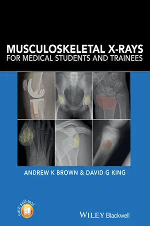
Musculoskeletal X-rays for Medical Students provides the key principles and skills needed for the assessment of normal and abnormal musculoskeletal radiographs. With a focus on concise information and clear visual presentation, it uses a unique colour overlay system to clearly present abnormalities.
Musculoskeletal X-rays for Medical Students:
- Presents each radiograph twice, side by side – once as would be seen in a clinical setting and again with clearly highlighted anatomy or pathology
- Focuses on radiographic appearances and abnormalities seen in common clinical presentations, highlighting key learning points relevant to each condition
- Covers introductory principles, normal anatomy and common pathologies, in addition to disease-specific sections covering adult and paediatric practice
- Includes self-assessment to test knowledge and presentation techniques
Musculoskeletal X-rays for Medical Students is designed for medical students, junior doctors, nurses and radiographers, and is ideal for both study and clinical reference.
Frequently asked questions
Yes, you can cancel anytime from the Subscription tab in your account settings on the Perlego website. Your subscription will stay active until the end of your current billing period. Learn how to cancel your subscription.
No, books cannot be downloaded as external files, such as PDFs, for use outside of Perlego. However, you can download books within the Perlego app for offline reading on mobile or tablet. Learn more here.
Perlego offers two plans: Essential and Complete
- Essential is ideal for learners and professionals who enjoy exploring a wide range of subjects. Access the Essential Library with 800,000+ trusted titles and best-sellers across business, personal growth, and the humanities. Includes unlimited reading time and Standard Read Aloud voice.
- Complete: Perfect for advanced learners and researchers needing full, unrestricted access. Unlock 1.4M+ books across hundreds of subjects, including academic and specialized titles. The Complete Plan also includes advanced features like Premium Read Aloud and Research Assistant.
We are an online textbook subscription service, where you can get access to an entire online library for less than the price of a single book per month. With over 1 million books across 1000+ topics, we’ve got you covered! Learn more here.
Look out for the read-aloud symbol on your next book to see if you can listen to it. The read-aloud tool reads text aloud for you, highlighting the text as it is being read. You can pause it, speed it up and slow it down. Learn more here.
Yes! You can use the Perlego app on both iOS or Android devices to read anytime, anywhere — even offline. Perfect for commutes or when you’re on the go.
Please note we cannot support devices running on iOS 13 and Android 7 or earlier. Learn more about using the app.
Yes, you can access Musculoskeletal X-Rays for Medical Students and Trainees by Andrew Brown,David G. King,Andrew K. Brown in PDF and/or ePUB format, as well as other popular books in Medicine & Medical Theory, Practice & Reference. We have over one million books available in our catalogue for you to explore.
Information
PART 1
Introduction
1
Musculoskeletal X‐rays
Introduction
Despite the availability of a wide range of imaging techniques to visualise musculoskeletal structures, plain radiographs or X‐rays remain an important and widely used first‐line investigation. It is probably the most readily available and least expensive imaging modality and is easily accessible to patients and healthcare professionals. As such, all medical professionals require at least a basic level of knowledge and training in the fundamental principles of requesting, interpreting and reporting plain radiographs of bones and joints.
Basic principles of requesting plain radiographs of bones and joints
Which structures?
Before requesting an X‐ray, the clinician should already have a good idea of the likely nature of the clinical problem and which musculoskeletal structures may be affected and what may or may not be visualised on an X‐ray. For example, radiographs are best at detecting pathological changes in bones, joints and cartilage, such as joint space narrowing, fracture, subluxation and dislocation. However, many soft tissue structures such as ligaments, tendons and synovium are not well visualised, and alternative imaging may be more appropriate to provide additional useful information to aid diagnosis and treatment.
Clinical assessment including the patient’s symptoms and a physical examination will usually determine the site or region that is to be evaluated by X‐ray. Exceptions may include a patient with rheumatoid arthritis where X‐rays of both hands and feet may be used to evaluate the extent of disease or structural damage which may be repeated serially to compare any disease progression over time.
Which views?
It is important to consider which views are chosen to visualise a particular region of interest. This is because the X‐ray beam creates a two dimensional shadow of a structure so selecting the correct view will maximise sensitivity. A basic principle is that two views should be requested, ideally 90° apart, which are usually obtained in antero‐posterior (AP) and lateral or oblique views. This is particularly important in a trauma context where fracture or dislocation may be missed if only a single view is acquired but may be less important for the assessment of arthritis. There are a number of established techniques and protocols for optimal image acquisition using standardised views of most musculoskeletal sites. Specialist views may also be considered in particular clinical situations, such as a ‘skyline view’ for the patello‐femoral joint or ‘through‐mouth view’ for the cervical spine. As well as the particular plane used to acquire the radiographic image, the position of the patient should also be considered. For example, weight‐bearing views with...
Table of contents
- Cover
- Title Page
- Table of Contents
- Preface
- Acknowledgements
- PART 1: Introduction
- PART 2: Pathology
- PART 3
- Self-assessment questions
- Index
- End User License Agreement