
Practical Manual of Echocardiography in the Urgent Setting
- English
- ePUB (mobile friendly)
- Available on iOS & Android
Practical Manual of Echocardiography in the Urgent Setting
About this book
Practical Manual of Echocardiography in the Urgent Setting
In the acute care setting, medicine happens at full speed and with little margin for error. As echocardiography plays an ever more important role in the diagnosis of patients who present with symptoms that suggest a cardiovascular emergency, clinicians must learn to collect, process and act on echocardiographic information as quickly and effectively as possible.
Practical Manual of Echocardiography in the Urgent Setting covers the essentials of echocardiography in the acute setting, from ultrasound basics to descriptions of all pertinent echocardiographic views to clear, stepwise advice on basic calculations and normal/abnormal ranges.
This compact new reference:
- Provides step-by-step guidance to acquiring the correct views and making the necessary calculations to accurately diagnose cardiac conditions commonly encountered in urgent settings.
- Presents information organized by complaint/initial presentation so that readers can work from this first knowledge of the patient through the steps required to pinpoint a diagnosis.
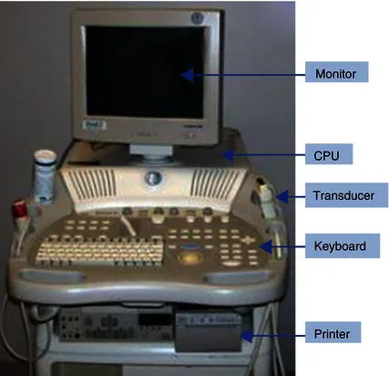
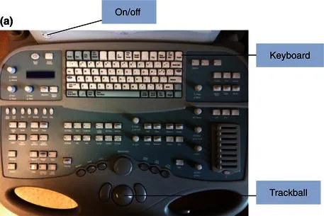
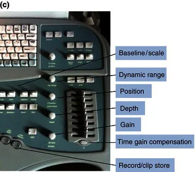
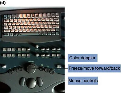
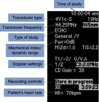
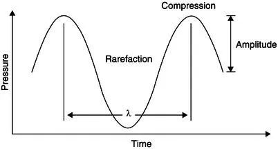
- Covers echo basics, from sound wave characteristics/properties to common device settings to basic ultrasound formulas
- Includes diagnostic algorithms fitted to address the differential diagnosis in the most commonlyencountered clinical scenarios.
Designed and written by frontline clinicians with extensive experience treating patients, Practical Manual of Echocardiography in the Urgent Setting is the perfect pocket-sized guide for residents in cardiology, emergency medicine, and hospital medicine; trainees in echocardiography; medical students on cardiology or emergency medicine rotations; technicians, nurses, attending physicians—anyone who practices in the urgent setting and who needs reliable guidance on echocardiographic views, data and normal/abnormal ranges to aid rapid diagnosis and decision-making at the point of care.
RELATED TITLES:
Kacharava, et al: Pocket Guide to Echocardiography; ISBN: 978-0-470-67444-4
Sun, et al: Practical Handbook of Echocardiography: 101 Case Studies; ISBN: 978-1-4051-9556-0
Frequently asked questions
- Essential is ideal for learners and professionals who enjoy exploring a wide range of subjects. Access the Essential Library with 800,000+ trusted titles and best-sellers across business, personal growth, and the humanities. Includes unlimited reading time and Standard Read Aloud voice.
- Complete: Perfect for advanced learners and researchers needing full, unrestricted access. Unlock 1.4M+ books across hundreds of subjects, including academic and specialized titles. The Complete Plan also includes advanced features like Premium Read Aloud and Research Assistant.
Please note we cannot support devices running on iOS 13 and Android 7 or earlier. Learn more about using the app.
Information
CHAPTER 1
Ultrasound physics


- M-mode: a graphic representation of a specific line of interest of a two-dimensional image (Figure 1.4).
- 2D: a two-dimensional view of cardiac structures that can be visualized as time progresses (Figure 1.5).
- Color Doppler: a color representation of blood flow velocities superimposed on a two-dimensional image (Figure 1.6).
- CW/PW Doppler: the representation of flow velocities as plotted with time on the x axis and velocity on the y axis (Figure 1.7).
- Tissue Doppler: the measurement of tissue velocities (Figure 1.8).


Ultrasound generation
- Cycle – the sum of one compression and one expansion of a sound wave.
- Frequency (f) – the number of cycles per second.
- Wavelength (λ) – the length of one complete cycle of sound.
- Period (p) – the time duration of one cycle.
- Amplitude – the maximum pressure change from baseline of a sound wave.
- Velocity (v) – speed at which sound moves through a specific medium.

Table of contents
- Cover
- Series page
- Title page
- Copyright page
- Contributors
- Preface
- CHAPTER 1: Ultrasound physics
- CHAPTER 2: The transthoracic examination
- CHAPTER 3: Transesophageal echocardiography
- CHAPTER 4: Ventricles
- CHAPTER 5: Left-sided heart valves
- CHAPTER 6: Right-sided heart valves
- CHAPTER 7: Prosthetic heart valves
- CHAPTER 8: The great vessels
- CHAPTER 9: Evaluation of the pericardium
- CHAPTER 10: Specialty echocardiographic examinations
- CHAPTER 11: Common artifacts
- CHAPTER 12: Hypotension and shock
- CHAPTER 13: Chest pain syndrome
- CHAPTER 14: Cardiac causes of syncope and acute neurological events
- CHAPTER 15: Acute dyspnea and heart failure
- CHAPTER 16: Evaluation of a new heart murmur
- CHAPTER 17: Infective endocarditis
- CHAPTER 18: Post-procedural complications
- CHAPTER 19: “Quick echo in the emergency department”: What the EM physician needs to know and do
- Index