Handbook of Microalgal Culture
eBook - ePub

Handbook of Microalgal Culture

Applied Phycology and Biotechnology

Amos Richmond, Qiang Hu

Buch teilen
  1. English
  2. ePUB (handyfreundlich)
  3. Über iOS und Android verfügbar
eBook - ePub

Handbook of Microalgal Culture

Applied Phycology and Biotechnology

Amos Richmond, Qiang Hu

Angaben zum Buch
Buchvorschau
Inhaltsverzeichnis
Quellenangaben

Über dieses Buch

Algae are some of the fastest growing organisms in the world, with up to 90% of their weight made up from carbohydrate, protein and oil. As well as these macromolecules, microalgae are also rich in other high-value compounds, such as vitamins, pigments, and biologically active compounds, All these compounds can be extracted for use by the cosmetics, pharmaceutical, nutraceutical, and food industries, and the algae itself can be used for feeding of livestock, in particular fish, where on-going research is dedicated to increasing the percentage of fish and shellfish feed not derived from fish meal. Microalgae are also applied to wastewater bioremediation and carbon capture from industrial flue gases, and can be used as organic fertilizer.

So far, only a few species of microalgae, including cyanobacteria, are under mass cultivation. The potential for expansion is enormous, considering the existing hundreds of thousands of species and subspecies, in which a large gene-pool offers a significant potential for many new producers.

Completely revised, updated and expanded, and with the inclusion of new Editor, Qiang Hu of Arizona State University, the second edition of this extremely important book contains 37 chapters. Nineteen of these chapters are written by new authors, introducing many advanced and emerging technologies and applications such as novel photobioreactors, mass cultivation of oil-bearing microalgae for biofuels, exploration of naturally occurring and genetically engineered microalgae as cell factories for high-value chemicals, and techno-economic analysis of microalgal mass culture. This excellent new edition also contains details of the biology and large-scale culture of several economically important and newly-exploited microalgae, including Botryococcus, Chlamydomonas, Nannochloropsis, Nostoc, Chlorella, Spirulina, Haematococcus, and Dunaniella species/strains.

Edited by Amos Richmond and Qiang Hu, each with a huge wealth of experience in microalgae, its culture, and biotechnology, and drawing together contributions from experts around the globe, this thorough and comprehensive new edition is an essential purchase for all those involved with microalgae, their culture, processing and use. Biotechnologists, bioengineers, phycologists, pharmaceutical, biofuel and fish-feed industry personnel and biological scientists and students will all find a vast amount of cutting-edge information within this Second Edition. Libraries in all universities where biological sciences, biotechnology and aquaculture are studied and taught should all have copies of this landmark new edition on their shelves.

Häufig gestellte Fragen

Wie kann ich mein Abo kündigen?
Gehe einfach zum Kontobereich in den Einstellungen und klicke auf „Abo kündigen“ – ganz einfach. Nachdem du gekündigt hast, bleibt deine Mitgliedschaft für den verbleibenden Abozeitraum, den du bereits bezahlt hast, aktiv. Mehr Informationen hier.
(Wie) Kann ich Bücher herunterladen?
Derzeit stehen all unsere auf Mobilgeräte reagierenden ePub-Bücher zum Download über die App zur Verfügung. Die meisten unserer PDFs stehen ebenfalls zum Download bereit; wir arbeiten daran, auch die übrigen PDFs zum Download anzubieten, bei denen dies aktuell noch nicht möglich ist. Weitere Informationen hier.
Welcher Unterschied besteht bei den Preisen zwischen den Aboplänen?
Mit beiden Aboplänen erhältst du vollen Zugang zur Bibliothek und allen Funktionen von Perlego. Die einzigen Unterschiede bestehen im Preis und dem Abozeitraum: Mit dem Jahresabo sparst du auf 12 Monate gerechnet im Vergleich zum Monatsabo rund 30 %.
Was ist Perlego?
Wir sind ein Online-Abodienst für Lehrbücher, bei dem du für weniger als den Preis eines einzelnen Buches pro Monat Zugang zu einer ganzen Online-Bibliothek erhältst. Mit über 1 Million Büchern zu über 1.000 verschiedenen Themen haben wir bestimmt alles, was du brauchst! Weitere Informationen hier.
Unterstützt Perlego Text-zu-Sprache?
Achte auf das Symbol zum Vorlesen in deinem nächsten Buch, um zu sehen, ob du es dir auch anhören kannst. Bei diesem Tool wird dir Text laut vorgelesen, wobei der Text beim Vorlesen auch grafisch hervorgehoben wird. Du kannst das Vorlesen jederzeit anhalten, beschleunigen und verlangsamen. Weitere Informationen hier.
Ist Handbook of Microalgal Culture als Online-PDF/ePub verfügbar?
Ja, du hast Zugang zu Handbook of Microalgal Culture von Amos Richmond, Qiang Hu im PDF- und/oder ePub-Format sowie zu anderen beliebten Büchern aus Technik & Maschinenbau & Fischerei & Aquakultur. Aus unserem Katalog stehen dir über 1 Million Bücher zur Verfügung.

Information

Jahr
2013
ISBN
9781118567197
Part 1
The Microalgal Cell with Reference to Mass Cultures
1
The Microalgal Cell
Robert A. Andersen
Friday Harbor Laboratories, University of Washington, Friday Harbor, WA, USA
Provasoli-Guillard National Center for Marine Algae and Microbiota, Bigelow Laboratory for Ocean Sciences, East Boothbay, ME, USA
Abstract
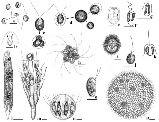
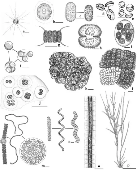
Microalgae are a diverse collection of microorganisms that conduct oxygen-evolving photosynthesis. Their biochemical diversity includes production of a wide array of carbohydrates, lipids, and proteins that are commercially valuable. Many produce several different morphologies, for example, flagellate, coccoid, and cyst stages. Many species are capable of sexual reproduction, some microalgae apparently having only asexual reproduction (e.g., Chlorella, Nannochloropsis). Algal ultrastructure is also diverse, paralleling their biochemical and physiological diversity. Many genomes of microalgae have been sequenced, and these are providing new insights into algal diversity. Genomic research has corroborated known endosymbiotic events and has revealed unknown, or cryptic, such events. Endosymbiosis has been a major factor in the production of algal diversity, and once it is better understood, this may be a practical means for producing new combinations of traits that have commercial application. The current state of algal taxonomy is summarized.
keywords algae; carbohydrate; chloroplast; endosymbiosis; genome; lipid; morphology; physiology; phytoplankton; protein
1.1 INTRODUCTION
Algae are primarily oxygen-releasing photosynthetic organisms with simple body plans – no roots, stems, or leaves. Algae are usually aquatic organisms. They do not form a single monophyletic group and consequently cannot be easily defined. Although algae as a group are ubiquitous, individual species occupy specific habitats. Some algae are attached to a substrate like plants, some are motile like animals, some are simply suspended in water, some grow loosely on soil, trees, and animals, and some form symbiotic relationships with other organisms (e.g., corals, lichens). The internal cell structure of algae varies greatly. Microalgae lack complex multicellular structures that are found in seaweeds. The cyanobacteria or blue-green algae have a prokaryotic cell structure and closely resemble bacteria. Eukaryotic algal cells have a nucleus and usually one or more chloroplasts; they also have mitochondria, Golgi bodies, endoplasmic reticulum, and other typical eukaryotic organelles. Despite the difficulty in presenting a clear definition for algae, thousands of books, scores of scientific journals, and numerous internet websites are dedicated solely to compiling our knowledge of algae (Lee, 2008; Graham et al., 2009).
1.2 GROSS MORPHOLOGY
Microalgae appear in a wide variety of shapes and forms. This morphological variation occurs not only among species but also among different life stages of the same species. The common forms are defined with adjectives such as amoeboid, palmelloid (= capsoid), coccoid, filamentous, flagellate, and sarcinoid (Figs. 1.1 and 1.2). Scientists use morphological life forms when generally discussing algae and their stages; there are, however, hundreds of thousands of algal species, and they do not always fit neatly into a few convenient categories. The first algae were morphologically simple organisms; today's simplest morphologies, however, are frequently the result of evolutionary reduction through which the algae are better able to survive because of their simplicity. In the following text, algal forms are treated from simple to complex, and this approach is strictly arbitrary (i.e., it does not reflect “primitive” vs. “advanced”).
Figure 1.1 Flagellate algal diversity. (a) Pedinomonas, with one visible flagellum. Scale bar = 5 μm (from Skuja, 1956). (b) Dunaliella, with two equal flagella. Scale bar = 10 μm (from Bold & Wynne, 1985). (c) Chlamydomonas, showing the biflagellate cell and four nonflagellate cells. Scale bar = 10 μm (flagellate from Ettl, 1976; colony from Skuja, 1956). (d) Haematococcus, showing the flagellate cell and three nonflagellate cells. Scale bar = 10 μm (from Skuja, 1948). (e) Tetraselmis, a quadraflagellate marine alga. Scale bar = 5 μm (from Throndsen, 1993). (f) Pavlova, with two unequal flagella and a very short haptonema. Scale bar = 5 μm (after Throndsen, 1993). (g) Isochrysis, with two nearly equal flagella and two chloroplasts. Scale bar = 5 μm (after Throndsen, 1993). (h) Synura, a colony with cells attached in the center. Scale bar = 10 μm (from Skuja, 1956). (i) Gymnodinium, a dinoflagellate with a circling transverse flagellum and a trailing longitudinal flagellum. Scale bar = 25 μm (from Skuja, 1956). (j) Ochromonas, with two very unequal flagella. Scale bar = 10 μm (from Skuja, 1964). (k) Chrysochromulina, with a long haptonema arising between the two flagella. Scale bar = 5 μm (after Throndsen, 1993). (l) Euglena terricola, a large cell with one flagellum emerging from a gullet. Scale bar = 10 μm (from Skuja, 1956). (m) Dinobryon, an arbuscular colony formed from loricas that surround each cell. Scale bar = 10 μm (from Skuja, 1964). (n) Stephanosphaera, a colony where cells are attached laterally. Scale bar = 10 μm (from Skuja, 1956). (o) Rhodomonas, a common marine biflagellate. Scale bar =10 μm (from Skuja, 1948). (p) Volvox, a large colonial flagellate with reproductive cells inside the otherwise hollow colony. Scale bar = 35 μm (from West, 1904).
c01f001
Figure 1.2 Diversity of nonflagellate algae. (a) Chrysamoeba mikrokonta, an amoeba with branching pseudopods. Scale bar = 5 μm (from Skuja, 1956). (b) Porphyridium purpureum, a single-celled red alga with a stellate chloroplast. Scale bar = 5 μm (from Hori, 1993b). (c) Synechococcus aeruginosus, the large freshwater type species of this cyanobacterium. Scale bar = 10 μm (from Geitler, 1932). (d) Nannochloropsis salina showing three elongate coccoid cells. Scale bar = 2 μm (from Andersen et al., 1998). (e) Nannochloropsis oculata showing four spherical coccoid cells. Scale bar = 2 μm (from Andersen et al., 1998). (f) Chlorella vulgaris showing a large single cell (top), four autospores (bottom), release of autospores (right). Scale bar = 10 μm (from Fott, 1959). (g) Scenedesmus maximus, showing four laterally connected coccoid cells. Scale bar = 10 μm (from Skuja, 1949). (h) Cosmarium ornatum, showing the typical semi-cell construction of desmids. Scale bar = 10 μm (from Skuja, 1956). (i) Oocystis gigas var. incrassata showing eight cells within the old mother cell wall. Scale bar = 20 μm (from Skuja, 1964). (j) Phacomyxa sphagnicola, a palmelloid alga showing vegetative cells within a colonial gelatinous matrix. Scale bar = 40 μm (from Skuja, 1956). (k) Botryococcus braunii showing cells in packets and numerous oil droplets in each cell. Scale bar = 10 μm (original). (l) Chlorosarcina superba showing a cuboidal colony. Scale bar = 10 μm (from Skuja, 1956). (m) Nostoc planctonicum showing an enlarged trichome (left), a long trichome, and the colony of trichomes. Scale bar = 5 μm (left, cells), = 25 μm (center, trichome), = 33 μm (colony) (from Geitler, 1932). (n) Spirulina/Arthrospira, showing different morphological forms of the spiraling trichome. Scale bar = 10 μm (left), = 18 μm (center), = 5 μm (right) (from Geitler, 1932). (o) Ulothrix moniliformis, an unbranched filament with a well-defined gelatinous sheath. Scale bar = 10 μm (from Skuja, 1956). (p) Cladophora sterrocladia, showing a typical branched filament shape. Scale bar = 250 μm (from Skuja, 1949).
c01f002
Flagellates may be single cells where each cell is an independent organism propelled through water with one or more flagella (e.g., Pedinomonas, Chlamydomonas, Gymnodinium, Ochromonas, Tetraselmis) (Fig. 1.1). Several to many flagellate cells may be joined together to produce a motile colony (e.g., Dinobryon, Synura). Large colonies, such as Volvox (Fig. 1.1p), have hundreds of cells. Most flagellate cells have two flagella, but marine picoflagellates may have only one flagellum (e.g., Micromonas and Pelagomonas) while Pyramimonas may have up to 16 flagella per cell. Haptophyte algae usually have a haptonema positioned between the two flagella (Fig. 1.1k), and the haptonema can be used for attaching to surfaces or collecting particles of food. Many common flagellate algae also produce nonmotile stages, as shown for Chlamydomonas and Haematococcus (Figs. 1.1c and 1.1d). Changing the environmental conditions can induce these alternate stages, and the manipulation of stages can be used to advantage in commercial facilities.
Many microalgae have a nonmotile stage as the dominant life form, and in some cases, no motile cells are ever found in the life cycle (Fig. 1.2). Amoeboid algae (e.g., Chlorarachnion, Chrysamoeba, Rhizochromulina) slowly creep across substrates, including the marine snow particles in oceans (Fig. 1.2a). Amoeboid cells may capture bacteria using pseudopods. Coccoid algae reproduce by autospores or zoospores, that is, mother cells undergo synchronized mitotic divisions and the number of daughter cells is fixed (e.g., 2, 4, 8, 16, 32). Single cells, such as Nannochloropsis are free, but commonly coccoid algae produce colonies (e.g., Chlorella, Oocystis, Scenedesmus) (Figs. 1.2d–g and 1.2i). Some, such as Synechococcus (Fig. 1.2c), exist today as single cells or weakly connected cells, but their ancestors were filamentous algae. Palmelloid algae have cells embedded within a gelatinous matrix; usually the cells are not physically connected to each other and only the gel holds them together. The gelatinous mass may be planktonic or attached to a substrate (Fig. 1.2j). The common flagellate Pavlova (Fig. 1.1f), for example, produces large palmelloid sheets when grown under certain culture conditions. Sarcinoid colonies result from equal cell division in three planes so that a cube is produced (Chlorosarcina; Fig. 1.2l). The oil-producing Botryococcus makes a crudely parenchymatous colony (Fig. 1.2k). Filaments are produced when cells attach end to end and form ribbon-like or chain-like assemblages. In their simplest form, filaments are unbranched and consist of a single row of cells (uniseriate) such as Arthrospira/Spirulina (Fig. 1.2n). Complexity develops with side branches (Fig. 1.2p) and multiple rows of cells (multiseriate). The cyanobacterium Nostoc forms large colonies that consist o...

Inhaltsverzeichnis