Small Animal Ophthalmology
eBook - ePub

Small Animal Ophthalmology

What's Your Diagnosis?

Heidi Featherstone, Elaine Holt

Buch teilen
  1. English
  2. ePUB (handyfreundlich)
  3. Über iOS und Android verfügbar
eBook - ePub

Small Animal Ophthalmology

What's Your Diagnosis?

Heidi Featherstone, Elaine Holt

Angaben zum Buch
Buchvorschau
Inhaltsverzeichnis
Quellenangaben

Über dieses Buch

Small Animal Ophthalmology: What's Your Diagnosis? is one of the first books in an exciting new series that combines problem-based learning, case studies, and questions and answers. Designed specifically for veterinarians and students, the series aims to present material in a format to enhance critical thinking and understanding.

Adopting a case-based approach, chapters are built around common ophthalmic presentations and are directed by questions to test the reader's ability to interpret clinical history, ophthalmic photographs and diagnostic results in order to provide differential diagnoses, diagnostic plans and treatment options.

For veterinary students, this book is an ideal guide to how ophthalmology cases are handled in the clinical setting. For veterinary practitioners, it is an innovative and interesting way to increase their knowledge and skills in clinical ophthalmology.

Häufig gestellte Fragen

Wie kann ich mein Abo kündigen?
Gehe einfach zum Kontobereich in den Einstellungen und klicke auf „Abo kündigen“ – ganz einfach. Nachdem du gekündigt hast, bleibt deine Mitgliedschaft für den verbleibenden Abozeitraum, den du bereits bezahlt hast, aktiv. Mehr Informationen hier.
(Wie) Kann ich Bücher herunterladen?
Derzeit stehen all unsere auf Mobilgeräte reagierenden ePub-Bücher zum Download über die App zur Verfügung. Die meisten unserer PDFs stehen ebenfalls zum Download bereit; wir arbeiten daran, auch die übrigen PDFs zum Download anzubieten, bei denen dies aktuell noch nicht möglich ist. Weitere Informationen hier.
Welcher Unterschied besteht bei den Preisen zwischen den Aboplänen?
Mit beiden Aboplänen erhältst du vollen Zugang zur Bibliothek und allen Funktionen von Perlego. Die einzigen Unterschiede bestehen im Preis und dem Abozeitraum: Mit dem Jahresabo sparst du auf 12 Monate gerechnet im Vergleich zum Monatsabo rund 30 %.
Was ist Perlego?
Wir sind ein Online-Abodienst für Lehrbücher, bei dem du für weniger als den Preis eines einzelnen Buches pro Monat Zugang zu einer ganzen Online-Bibliothek erhältst. Mit über 1 Million Büchern zu über 1.000 verschiedenen Themen haben wir bestimmt alles, was du brauchst! Weitere Informationen hier.
Unterstützt Perlego Text-zu-Sprache?
Achte auf das Symbol zum Vorlesen in deinem nächsten Buch, um zu sehen, ob du es dir auch anhören kannst. Bei diesem Tool wird dir Text laut vorgelesen, wobei der Text beim Vorlesen auch grafisch hervorgehoben wird. Du kannst das Vorlesen jederzeit anhalten, beschleunigen und verlangsamen. Weitere Informationen hier.
Ist Small Animal Ophthalmology als Online-PDF/ePub verfügbar?
Ja, du hast Zugang zu Small Animal Ophthalmology von Heidi Featherstone, Elaine Holt im PDF- und/oder ePub-Format sowie zu anderen beliebten Büchern aus Medizin & Veterinärmedizin. Aus unserem Katalog stehen dir über 1 Million Bücher zur Verfügung.

Information

Jahr
2011
ISBN
9781444340020
Auflage
1
Thema
Medizin
CHAPTER 1
Abnormalities of Globe Size and Position
Introduction
It can be a challenge to differentiate between a change in size and a change in position of the eye. An abnormally small eye (microphthalmos) may be confused with a normal-sized eye that is recessed in the orbit (enophthalmos); an enlarged eye (buphthalmos) may have a similar appearance to a normal-sized eye that is anteriorly displaced (exophthalmos). Assessing the size of the palpebral fissure, position of the third eyelid (TEL) and corneal diameter, looking at the eye from different angles, careful comparison with the other eye and concurrent clinical signs are helpful in differentiating between these conditions.
CASE STUDY 1
History
A 12-year-old female neutered domestic shorthaired cat is presented because of a sudden redness in the right eye. The left eye had looked abnormal for several weeks but appeared comfortable. The cat has recently lost weight and is lethargic.
Questions
1. Describe the abnormalities in Figs. 1.1a, b, and c.
Fig. 1.1a
c01f001a
Fig. 1.1b
c01f001b
Fig. 1.1c
c01f001c
2. What differential diagnoses should be considered for this presentation?
3. What tests could you perform to make the diagnosis?
Answers
1. What the Figures Show
Fig. 1.1a The left eye appears larger than the right eye; a wide palpebral fissure, increased corneal diameter and clearly visible medial and lateral regions of the limbus are consistent with buphthalmos. There is a generalised corneal opacity which is most dense axially; a tapetal reflection is not visible. In the right eye, the green tapetal reflection is obstructed ventrally by a red/black irregular opacity which appears to be in front of the iris, and there is a similar coloured opacity overlying the iris at the 9 o’clock position. The pupil is moderately dilated.
Fig. 1.1b In the left eye, the Purkinje images are disrupted. There is generalised corneal vascularisation and a stippled area of fluorescein stain uptake axially. The conjunctival vessels overlying the sclera on the lateral aspect of the globe are congested. The iris is difficult to see well but appears darker (medially) and possibly thickened. In the right eye, there is hyphaema; the regions of the iris that are visible appear normal.
Fig. 1.1c Oblique view from the lateral aspect of both eyes. In the left eye there is an irregular contour and anterior protrusion of the cornea (OS > OD). There is increased exposure of the sclera and conjunctiva, and episcleral congestion. The anterior chamber is obliterated by abnormal iris tissue which appears to be displaced anteriorly. In both eyes fluorescein dye is visible on the periocular hair at the medial canthus.
2. Differential Diagnoses
Given the history and the appearance of the left eye, the following conditions should be considered:
  • Chronic glaucoma In contrast to the dog, primary glaucoma in the cat is rare, and secondary glaucoma is more common. The most common causes of secondary glaucoma in the cat are chronic idiopathic lymphocytic-plasmacytic uveitis and primary intraocular neoplasia, most notably diffuse iris melanoma. Typical clinical signs include buphthalmos, conjunctival and episcleral congestion, corneal oedema, mydriasis, and impaired or absent vision. Buphthalmos can be difficult to discern in the cat and assessment of the size of the palpebral fissure can be helpful because it becomes wider as the size of the eye increases. Glaucoma in cats is typically insidious in onset and is often difficult to recognise. This is in contrast to canine primary glaucoma which is characterised by peracute pain, episcleral congestion, marked corneal oedema, mydriasis and blindness (Ch. 6, case 2).
  • Exophthalmos Anterior displacement of the globe within the orbit. Common causes of exophthalmos in the cat include orbital neoplasia, orbital cellulitis/abscess and orbital trauma (haematoma, emphysema, fracture, foreign body). Primary malignant neoplasia and abscesses secondary to dental disease are more likely in old cats, whereas head trauma and orbital foreign bodies are more common in young cats (Ch.12, case 2).
Given the appearance of the right eye, the following conditions should be considered:
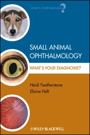
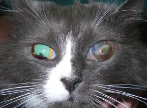
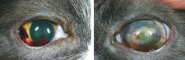
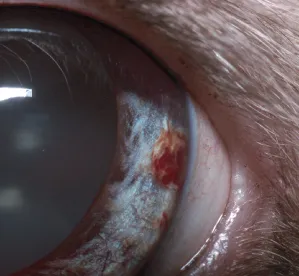
  • Systemic hypertension Sustained systemic hypertension is commonly associated with ocular manifestations which primarily involve the posterior segment but also affect the anterior segment. Abnormalities in the posterior segment involve the retina, choroid and vitreous humour and appear as retinal oedema and bullae, retinal and intravitreal haemorrhages, retinal detachment and increased tortuosity of the retinal arterioles. Intraocular haemorrhage can occur as a result of haemorrhage from the iris (Fig. 1.1d), ciliary body, retina, and choroid. Extensive hyphaema can lead to the formation of anterior and posterior synechiae and secondary glaucoma.
  • Coagulopathy and platelet disorders Ocular haemorrhage can be a clinical sign of a coagulopathy or a platelet disorder. Ocular haemorrhage typically occurs when the platelet count is <50 000 cells/µl.
  • Uveitis When there is a breakdown of the blood-aqueous barrier during inflammation, red blood cells can enter the anterior chamber (hyphaema). The blood may form either a homogenous layer in the ventral anterior chamber or a clot, as in this cat.
  • Trauma Ocular haemorrhage may result from both blunt and penetrating ocular trauma (Ch. 12, cases 2 and 3).
  • Pre-iridal fibrovascular membrane (PIFM) The formation of fibrovascular membranes on the anterior iris is usually a consequence of intraocular inflammation, haemorrhage and/or hypoxia due to the release of vasoactive substances. Hence the formation of PIFMs is common in eyes with chronic uveitis, intraocular haemorrhage, retinal detachment, glaucoma, and neoplasia. The newly formed blood vessels within the membranes are fragile and can cause spontaneous and recurrent hyphaema. PIFMs can extend into the filtration angle and result in secondary glaucoma. Fibrovascular membranes are not restricted to the surface of the iris – they can also form on the retina and optic disc and in the vitreous.
  • Neoplasia (primary or secondary) Intraocular haemorrhage may occur in eyes affected with primary or secondary neoplasia, either originating from a PIFM or as a result of the direct effect of neoplasia (e.g. adverse effect on clotting function).
  • Congenital anomalies These include persistent hyaloid artery and persistent hyperplastic primary vitreous, both of which are rare conditions in the cat.
Fig. 1.1d Aneurysm within the lateral region of the major arterial circle in the left eye of a cat with systemic hypertension.
c01f001d
3. Appropriate Diagnostic Tests
  • Ocular reflexes
    • Pupillary light reflex – the left pupil is not visible. Negative consensual OS (from left to right eye); positive direct OD, albeit slow and incomplete.
    • Dazzle reflex – negative OS, positive OD
    • Palpebral reflex – positive OU, OS < OD
    • Corneal reflex – positive OU, OS < OD
  • Menace response – negative OS, equivocal OD
In this cat, these results are consistent with blindness, reduced corneal sensation and lagophthalmos in the left eye, and reduced vision in the right eye.
  • Examination with a focal light source – in the left eye, slit-lamp biomicroscopy reveals extensive superficial and deep corneal vascularisation, and generalised corneal oedema and fibrosis which is most marked axially.
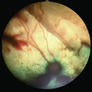
  • Ophthalmoscopy – in the right eye, this reveals an extensive dorsal retinal detachment, most marked within the medial quadrant, and multiple retinal haemorrhages of different sizes throughout the tapetal fundus and ventral to the optic disc (Fig. 1.1e).
  • Schirmer tear test – 4 mm/min OS, 10 mm/min OD
  • Fluorescein dye – negative staining OD, positive staining in the superficial axial cornea OS. This is indicative of suboptimal ocular surface health in the left eye, most likely because of the lagophthalmos.
  • Tonometry – IOP 35 mmHg OS, 20 mmHg OD
Fig. 1.1e Extensive dorsal retinal detachment and multiple retinal haemorrhages throughout the tapetal fundus and ventral to the optic disc.
c01f001e
There is increased resistance to retropulsion of the left eye; retropulsion of the right eye is normal. The remainder of the ophthalmic examination reveals no additional abnormalities. A general physical examination reveals an underweight body condition and mild dental disease.
The degree of resistance to retropulsion of the eye varies amongst species and between breeds. The normal feline globe is generally retropulsed less than the normal canine globe because of close apposition between the globe and the orbit in the cat. The degree of retropulsion in brachycephalic breeds is less than in other breeds because of the shallow orbit, in both cats and dogs.
Further Diagnostic Tests
  • B-mode ocular ultrasound – this is indicated to evaluate the posterior segment when the anterior segment is opaque, and to take m...

Inhaltsverzeichnis