
eBook - ePub
Atlas of Endoscopic Ultrasonography
Frank G. Gress, Thomas J. Savides, Brenna C. Bounds, John C. Deutsch, Frank G. Gress, Thomas J. Savides, Brenna C. Bounds, John C. Deutsch
This is a test
Buch teilen
- English
- ePUB (handyfreundlich)
- Über iOS und Android verfügbar
eBook - ePub
Atlas of Endoscopic Ultrasonography
Frank G. Gress, Thomas J. Savides, Brenna C. Bounds, John C. Deutsch, Frank G. Gress, Thomas J. Savides, Brenna C. Bounds, John C. Deutsch
Angaben zum Buch
Buchvorschau
Inhaltsverzeichnis
Quellenangaben
Über dieses Buch
The Atlas of Endoscopic Ultrasonography provides readers with a large collection of excellent images obtained from both diagnostic and therapeutic procedures. The Atlas includes a DVD which will be an invaluable addition to the library of trainee and practising gastroenterologists with video clips and searchable database of images. Together the book and DVD offer a first class collection of images to give a highly integrated introduction to endoscopic ultrasonography.
The Atlas is an ideal companion to Dr Gress et al's Endoscopic Ultrasonography, Second Edition.
Häufig gestellte Fragen
Wie kann ich mein Abo kündigen?
Gehe einfach zum Kontobereich in den Einstellungen und klicke auf „Abo kündigen“ – ganz einfach. Nachdem du gekündigt hast, bleibt deine Mitgliedschaft für den verbleibenden Abozeitraum, den du bereits bezahlt hast, aktiv. Mehr Informationen hier.
(Wie) Kann ich Bücher herunterladen?
Derzeit stehen all unsere auf Mobilgeräte reagierenden ePub-Bücher zum Download über die App zur Verfügung. Die meisten unserer PDFs stehen ebenfalls zum Download bereit; wir arbeiten daran, auch die übrigen PDFs zum Download anzubieten, bei denen dies aktuell noch nicht möglich ist. Weitere Informationen hier.
Welcher Unterschied besteht bei den Preisen zwischen den Aboplänen?
Mit beiden Aboplänen erhältst du vollen Zugang zur Bibliothek und allen Funktionen von Perlego. Die einzigen Unterschiede bestehen im Preis und dem Abozeitraum: Mit dem Jahresabo sparst du auf 12 Monate gerechnet im Vergleich zum Monatsabo rund 30 %.
Was ist Perlego?
Wir sind ein Online-Abodienst für Lehrbücher, bei dem du für weniger als den Preis eines einzelnen Buches pro Monat Zugang zu einer ganzen Online-Bibliothek erhältst. Mit über 1 Million Büchern zu über 1.000 verschiedenen Themen haben wir bestimmt alles, was du brauchst! Weitere Informationen hier.
Unterstützt Perlego Text-zu-Sprache?
Achte auf das Symbol zum Vorlesen in deinem nächsten Buch, um zu sehen, ob du es dir auch anhören kannst. Bei diesem Tool wird dir Text laut vorgelesen, wobei der Text beim Vorlesen auch grafisch hervorgehoben wird. Du kannst das Vorlesen jederzeit anhalten, beschleunigen und verlangsamen. Weitere Informationen hier.
Ist Atlas of Endoscopic Ultrasonography als Online-PDF/ePub verfügbar?
Ja, du hast Zugang zu Atlas of Endoscopic Ultrasonography von Frank G. Gress, Thomas J. Savides, Brenna C. Bounds, John C. Deutsch, Frank G. Gress, Thomas J. Savides, Brenna C. Bounds, John C. Deutsch im PDF- und/oder ePub-Format sowie zu anderen beliebten Büchern aus Medicine & Gastroenterology & Hepatology. Aus unserem Katalog stehen dir über 1 Million Bücher zur Verfügung.
Information
Part 1: Normal EUS Anatomy
1
Normal Human Anatomy
Introduction
The Visible Human Project at the University of Colorado has generated large volumes of human anatomy data. The original information is captured by slowly abrading away frozen human cadavers in a transaxial manner and capturing the anatomy by digital imaging. The digital data is compiled and then over the years is manipulated by scientists at the University’s Center for Human Simulation to allow access to identified cross sections in any plane as well as to models which can be lifted from the data set. Details regarding the Visible Human Project and its applications to gastroenterology and endosonography have been previously described.
This atlas is fortunate to be able to use the interactive anatomy resources developed by Vic Spitzer, Karl Reinig, David Rubenstein, and others to create movies that help explain what takes place during endoscopic ultrasound (EUS) evaluations. Since EUS is a “real-time” examination, it seems reasonable to present this section primarily as “real-time” videos. The videos can be viewed over and over, allowing endosonographers to look not only at the highlighted structures, but also at structures they might visualize during EUS that are not specifically identified on the selected video.
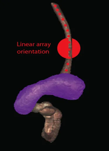
This chapter uses the terms “radial array orientation” to describe planar anatomy which would be found perpendicular to a line going through the digestive tract (as would be generated by a radial array echoendoscope, Figure 1.1) and “linear array orientation” for planar anatomy generated parallel to a line going through the digestive tract (as would be generated by a linear array echoendoscope, Figure 1.2).
Figure 1.1 Visible Human Model of esophagus, stomach, and duodenum. The green circle shows a plane perpendicular to the axis and is similar to a plane developed during radial array endosonography.
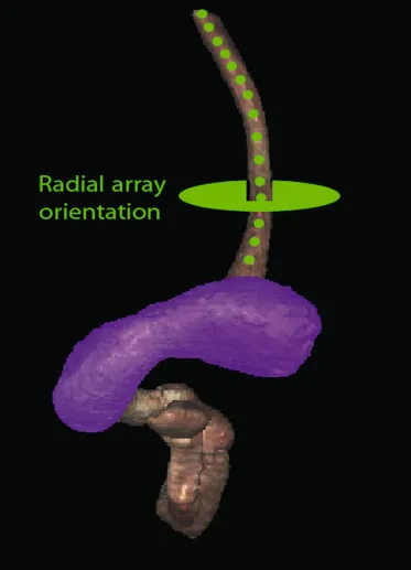
Figure 1.2 Visible Human Model of esophagus, stomach, and duodenum. The red circle shows a plane parallel to the axis and is similar to a plane developed during linear array endosonography.
Normal EUS Anatomy from the Esophagus
Radial Array Orientation (Video 1.1)
Video 1.1 starts with Visible Human Models of the left atrium (purple), trachea and bronchi (light blue), aorta and pulmonary arteries (red), vena cava (dark blue), and the esophagus (brown). A plane is shown passing through the esophagus. This plane contains the transaxial cross-sectional anatomy images which then follow, starting in the oropharynx and going caudally. The upper esophageal sphincter (UES) is identified. As the images proceed distally, the trachea and esophagus can be followed to a point where the brachiocephalicn left carotid and left subclavian arteries are evident just above the aortic arch. Below the aortic arch is the aortopulmonary window. The azygous arch can be seen exiting the superior vena cava (SVC). This occurs just above the tracheal bifurcation. The esophagus, labeled as “E” is surrounded by the descending aorta, the vertebrae, and the trachea. The thoracic duct (not labeled) is visible between the aorta and vertebrae, inferior to the esophagus. Going distally, the pulmonary artery becomes prominent. The region between the right mainstem bronchus (RMB) and left mainstem bronchus (LMB) is the subcarinal space. The video progresses to a level where the left artrium surrounds the superior aspect of the esophagus and then the video ends as the esophagus passes the gastroesophageal junction.
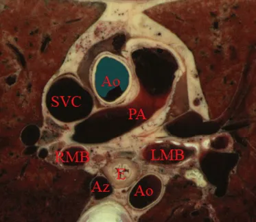
An image plane cross-section of taken from a radial array orientation at the level of the subcarinal space is shown (Figure 1.3).
Figure 1.3 Transaxial cross-section of digital anatomy taken at the level of the subcarinal space (Ao = aorta [both ascending, superior in the image, and descending, inferior in the image, are shown]; Az = azygos vein; PA = pulmonary artery; RMB and LMB = right and left main stem bronchi; SVC = superior vena cava).
Linear Array Orientation (Video 1.2)
Video 1.2 starts with the same models as above (The left atrium [purple], trachea and bronchi [light blue], aorta and pulmonary arteries [red], vena cava [dark blue], and the esophagus [brown]). The plane shows potential ways that cross-sectional anatomy can be generated. The video then shows a sagittal image with the descending aorta inferior to the esophagus, much as what is done during linear array EUS. In this orientation the pulmonary artery (PA) and left atrium are superior. The image plane is rotated to bring the left atrium and pulmonary artery to the inferior side of the esophagus. The models are then shown again, and the plane is moved in the caudal and cephalad directions, much as during EUS.
Normal EUS Anatomy from the Stomach
Radial Array Orientation (Video 1.3)
Endoscopic ultrasound of the stomach differs from EUS at other sites since the stomach does not constrain the endoscope tightly. It is important to follow anatomical structures (such as in a station approach) to avoid getting lost.
The video shows models of the stomach, esophagus, duodenum, gallbladder, pancreas (brown), the aorta, splenic artery, hepatic artery and left gastric artery (red), adrenal glands (pink), and splenic, superior mesenteric veins (dark blue) as viewed from behind. A plane is passed that is similar to the image plane generated during radial array EUS. The resultant cross-sectional anatomy starts at the level of the gastroesophageal junction, with the aorta and inferior vena cava (IVC) labeled. The aorta (which is collapsed) is followed, which brings the pancreas and left adrenal gland into view. The first artery that comes off the aorta in the abdomen is the celiac artery. There is a trifurcation into the splenic, hepatic, and left gastric arteries (LGA), although the LGA is generally smaller and difficult to see. It is shown in the video at the “x” just before the bifurcation into the celiac and hepatic arteries as identified.
The superior mesenteric artery (SMA) comes off the aorta just distal to the celiac artery. Various endoscope maneuvers can be used to bring the portal confluence into view, and then the splenic vein can be used as a guide to visualize the pancreas body, left adrenal, kidney, and spleen. The diaphragm can be easily imaged between the kidney and the vertebrae.
Linear Array Orientation (Video 1.4)
The linear array exam also follows the aorta to the stomach, but, as shown Video 1.4, the image plane across the pancreas is generally obtained through a sweeping motion. The first major gastric landmark is the origin of the celiac artery and SMA from the aorta (Figure 1.4). The superior mesenteric vein (SMV), portal vein, and splenic vein can be used as guides to go back and forth across the pancreas and in the process, the left adrenal, kidney, and spleen can be seen. The splenic artery runs roughly parallel to the splenic vein, but is generally tortuous.
Figure 1.4 Sagital cross-section of digital anatomy at the level of the gastroesophageal junction, similar to a view seen during linear array endoscopic ultrasound (EUS). The celiac and superior mesenteric arteries (SMA) are shown at their insertion into the aorta. The renal vein (RV) is shown adjacent to the SMA and the splenic vein is shown adjacent to the pan...