
eBook - ePub
Human Anatomy for Art Students
Ralph Thompson, Sir Alfred Fripp
This is a test
Buch teilen
- 352 Seiten
- English
- ePUB (handyfreundlich)
- Über iOS und Android verfügbar
eBook - ePub
Human Anatomy for Art Students
Ralph Thompson, Sir Alfred Fripp
Angaben zum Buch
Buchvorschau
Inhaltsverzeichnis
Quellenangaben
Über dieses Buch
A staple of art instruction, this book is the most concise and accessible guide to accurately depicting the human body. Its illustrations and cross-sections offer examples of human skeletal and muscular substructures and details of individual body parts, helping students achieve the most precise visual re-creation of human form and motion.
Subjects include the skeleton, the coverings and regions of the body, the upper and lower extremities, movements of the joints, the trunk, and the head and neck. Additional topics encompass expression and gesture, differences of size and proportion in the sexes, and growth, development, and measurements. More than 130 detailed figures appear throughout the text, in addition to thirty-one plates.
Subjects include the skeleton, the coverings and regions of the body, the upper and lower extremities, movements of the joints, the trunk, and the head and neck. Additional topics encompass expression and gesture, differences of size and proportion in the sexes, and growth, development, and measurements. More than 130 detailed figures appear throughout the text, in addition to thirty-one plates.
Häufig gestellte Fragen
Wie kann ich mein Abo kündigen?
Gehe einfach zum Kontobereich in den Einstellungen und klicke auf „Abo kündigen“ – ganz einfach. Nachdem du gekündigt hast, bleibt deine Mitgliedschaft für den verbleibenden Abozeitraum, den du bereits bezahlt hast, aktiv. Mehr Informationen hier.
(Wie) Kann ich Bücher herunterladen?
Derzeit stehen all unsere auf Mobilgeräte reagierenden ePub-Bücher zum Download über die App zur Verfügung. Die meisten unserer PDFs stehen ebenfalls zum Download bereit; wir arbeiten daran, auch die übrigen PDFs zum Download anzubieten, bei denen dies aktuell noch nicht möglich ist. Weitere Informationen hier.
Welcher Unterschied besteht bei den Preisen zwischen den Aboplänen?
Mit beiden Aboplänen erhältst du vollen Zugang zur Bibliothek und allen Funktionen von Perlego. Die einzigen Unterschiede bestehen im Preis und dem Abozeitraum: Mit dem Jahresabo sparst du auf 12 Monate gerechnet im Vergleich zum Monatsabo rund 30 %.
Was ist Perlego?
Wir sind ein Online-Abodienst für Lehrbücher, bei dem du für weniger als den Preis eines einzelnen Buches pro Monat Zugang zu einer ganzen Online-Bibliothek erhältst. Mit über 1 Million Büchern zu über 1.000 verschiedenen Themen haben wir bestimmt alles, was du brauchst! Weitere Informationen hier.
Unterstützt Perlego Text-zu-Sprache?
Achte auf das Symbol zum Vorlesen in deinem nächsten Buch, um zu sehen, ob du es dir auch anhören kannst. Bei diesem Tool wird dir Text laut vorgelesen, wobei der Text beim Vorlesen auch grafisch hervorgehoben wird. Du kannst das Vorlesen jederzeit anhalten, beschleunigen und verlangsamen. Weitere Informationen hier.
Ist Human Anatomy for Art Students als Online-PDF/ePub verfügbar?
Ja, du hast Zugang zu Human Anatomy for Art Students von Ralph Thompson, Sir Alfred Fripp im PDF- und/oder ePub-Format sowie zu anderen beliebten Büchern aus Art & Art Techniques. Aus unserem Katalog stehen dir über 1 Million Bücher zur Verfügung.
Information
Thema
ArtThema
Art TechniquesCHAPTER I
THE SKELETON
No bone is either quite rigid or quite straight. The elasticity (which is greater in the young) and the curves found in every bone are obviously adapted to increase its strength.
The skeleton of the adult is built up of almost rigid bones, and the length of each bone is slightly increased by a layer of less rigid cartilage, forming a kind of buffer at each end. In addition, within certain of the joints, actual cushions of fibro-cartilage intervene between the ends of the bones.
There are various forms of bone. The femur, or thigh-bone, may be taken as an example of the long bone, while the spine, wrist, and ankle are composed of short bones. The scapula, or shoulder-blade, and the frontal, or forehead bone, are good examples of the ƒlat bone.
The stature of any individual depends chiefly upon the length of the long bones of the lower limb and of the short bones of the spinal column.
1. The long bones should be regarded as mechanical “levers”; every muscular action may be interpreted as a power or force applied to such a part of the bar or bone as to overcome a definite weight or resistance, and so to produce movement about a fixed point or fulcrum.
2. The short bones will plainly be less liable to fracture, and the multiplication of the cartilaginous and articulating surfaces will, of course, result in the better breaking of jars and increased mobility. Thus, if the spine were a single rigid long bone, its relation to the cranium would be that of the broom-handle to the broom-head, and the effect of a blow upon the other end, as when one sits down with a jerk, would be to drive the neck into the base of the skull, just as the handle of the broom is driven into the broom-head.
3. The flat bones are generally protective; thus the flat bones of the vault of the skull protect the delicate brain which lies in the cranium, and the flat bones on each side of the pelvis, known as the hip-bones or ossa innominata, afford protection to important viscera. In addition, flat bones provide an extensive attachment for strong muscles. Chewing or mastication, which is one of the most powerful and one of the most fundamentally important movements in the body, is brought about by the very strong muscles inserted into the jaw, which have an extensive origin from the flat bones of the cranium. In like manner the movements of the humerus are partly caused by muscles which arise from broad areas of the scapula.
A muscle is said to “arise or take origin ” from that end which usually is fixed when the muscle acts, and its “insertion ” is that end which usually moves most.
The femur is the longest bone in the body; the next longest are the bones of the leg and arm and forearm, some of the ribs, and then the clavicle.
The bones of the upper limb comprise:—
- The clavicle, or collar-bone.
- The scapula, or shoulder-blade.
- The humerus, or bone of the arm.
- The ulna, the inner bone of the forearm.
- The radius, the outer bone of the forearm.
- The carpals or bones of the wrist.
- The metacarpals or bones of the palm of the hand.
- The phalanges or bones of the digits.
1. The clavicle (Fig. 1) is situated in the front part of the thorax or chest, where the trunk merges into the neck. At its inner extremity it is joined to the sternum (breast-bone) by the sterno-clavicular joint. The inner ends of the right and left clavicles are about an inch apart. The outer extremity of the clavicle touches the acromion process of the scapula in the acromio-clavicular joint, and is situated at a somewhat higher level than the inner end (Fig. 1). This is the case even with people whose shoulders slope in a very marked degree.
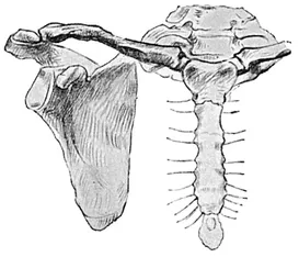
Fig. 1.—The Clavicle and Scapula of the Right Side, with the Sternum. From the front.
The clavicle has its curves so arranged that there is a convexity forward in the inner part, for rather more than half the length of the bone, and a concavity forward in the outer part, for rather less than half. It is thicker and more prominent internally than externally: a cross section made through the internal half is triangular; through the external half it is flattened from above downwards.
In the clavicle, as in all other long bones, the degree of its roughness gives a fair indication of the muscularity of the individual.
The clavicle is a bone of high importance to the student of anatomy. It forms a very prominent landmark, easily seen in thin people. In well-covered and muscular subjects, however, it lies at the bottom of a furrow, a situation common in many other parts of the body, e.g. the external condyle of the humerus and the great trochanter of the femur, a dimple indicating the position of a bone which in the skeleton appears to be prominent.
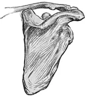
Fig. 2.—The Scapula and Clavicle of the Right Side. From the back.
The clavicle, unlike the other long bones, continues to increase much in length between the ages of twenty and twenty-five years, and thus produces, during this period, a great increase in the breadth of the shoulders, an increase which constitutes one of the chief characteristics distinguishing the adolescent boy from the adult man.
2. The scapula (Fig. 2) is chiefly visible upon the upper part of the back of the trunk, but two of its “ processes are apparent from the front of the skeleton, namely, the acromion, which makes the point of the shoulder, and the coracoid, which is covered by thick muscles, but in the wasted subject can be seen under the skin below the outer part of the clavicle.
It is a triangular flat bone having two sur. faces:—
a. The front or ventral surface, applied to the ribs over the back of the thorax, from the second to the seventh.
b. The hinder or dorsal surface, overlaid by the muscles and skin of the back.
Three borders:—
a. The upper.
b. The mesial, vertical, or vertebral.
c. The external, oblique, or axillary (lying in relation to the axilla or armpit).
And three angles:—
a. The superior.
b. The inferior, which is prominent in persons who are “round-shouldered.”
c. The external, sometimes called the head of the scapula, which, with the head of the humerus and the connecting ligaments, forms the shoulder-joint.
The three processes of the scapula are:—
a. The spine or spinous process (Fig. 2, p. 36). This is a well-marked bony ridge which projects backwards from the dorsal surface. It begins internally at the junction of the upper and second quarter of the vertebral border, and becomes more prominent externally, where it terminates in the second process.
b. The acromion process. This is flattened and directed forwards, upwards, and outwards, to form the point of the shoulder. The acromion process has two borders, of which the inner one enters, with the external end of the clavicle, into the formation of the acromio-clavicular joint.
c. The coracoid process is curved upon itself, and tapers rapidly to its apex, which is directed forwards and outwards just below the forward concavity of the outer third of the clavicle (Fig. 1).
3. The humerus (Fig. 3), or bone of the arm proper, is described, like all the long bones, as consisting of a shaft and two extremities, upper and lower. (The extremities of the clavicle only are known as outer and inner, and the extremities of the ribs are called anterior and posterior.)
The humerus articulates above with the head of the scapula, to form the shoulder-joint, and below with the ulna and radius, where it forms the elbow-joint.
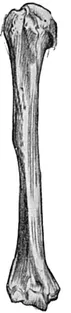
Fig. 3.—The Right Humerus. Front view.
The upper extremity, or head of the humerus, forms a small segment of a large globe. It is directed upwards, inwards, and backwards, and the size of it greatly influences the prominently convex shape and outline of the shoulder. When the arm is outstretched, the head of the humerus may be felt, or even seen, in the axilla, especially in thin people.
The tuberosities of the humerus are flattened projections which are separated from the head by the anatomical neck, to which the capsule of the shoulder-joint is attached. The vertical bicipital groove divides the greater and lesser tuberosities from each other, the groove running downwards, inwards, and slightly forward, and lodging the tendon of the long head of the biceps muscle. The greater tuberosity lies outside and behind the lesser.
The shaft of the humerus begins below the tuberosities and head at a decidedly narrower part, which is known as the surgical neck on account of the frequency with which the bone is broken in this region. Below the neck the shaft becomes a little thicker, and is twisted, not bent, outwards through 38 an angle of fifteen or twenty degrees. In its lower third it is slightly flattened from before backwards, and is concave forwards.
The lower extremity of the humerus is very wide from side to side.
The condyles of the humerus lie on each side of the lower extremity. The internal, which is larger and lies at a lower level than the external, is directed chiefly inwards but also slightly backwards, while the external is directed outwards.
The condyles serve the usual purpose of bony prominences, namely, the attachment of muscles in this case, and of some of the ligaments of the neighbouring joint, the elbow. They form the lower limits of corresponding ridges, called the supra-condylar ridges, which descend on each side of the shaft of the humerus.
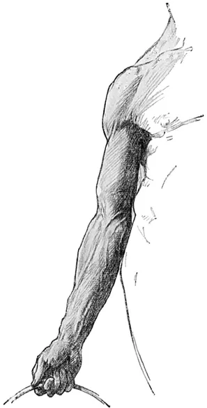
Fig. 4.—The Front of the Upper Extremity, showing the “Carrying Angle” between Arm and Forearm.
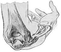
Fig 5.—The Bones forming the Elbow Joint. Back view.
The trochlea is a broad, smooth articular surface at the lower end of the humerus, grooved obliquely, so that behind the groove it is directed downwards and inwards, and in front it runs upwards and outwards. The inner lip of the trochlea is more prominent than the outer, especially towards its lower part. This lip, by its large size, is responsible for the maintenance of the carrying angle (Fig. 4). The articulation of ...