Temporary Anchorage Devices in Clinical Orthodontics
eBook - ePub

Temporary Anchorage Devices in Clinical Orthodontics

Jae Hyun Park, Jae Hyun Park

Buch teilen
  1. English
  2. ePUB (handyfreundlich)
  3. Über iOS und Android verfügbar
eBook - ePub

Temporary Anchorage Devices in Clinical Orthodontics

Jae Hyun Park, Jae Hyun Park

Angaben zum Buch
Buchvorschau
Inhaltsverzeichnis
Quellenangaben

Über dieses Buch

Provides the latest information on all aspects of using temporary anchorage devices in clinical orthodontics, from diagnosis and treatment planning to appliances and applications

Written by some of the world's leading experts in orthodontics, Temporary Anchorage Devices in Clinical Orthodontics is a comprehensive, up-to-date reference that covers all aspects of temporary anchorage device (TAD) use in contemporary orthodontics. Taking a real-world approach to the subject, it covers topics ranging from diagnosis and treatment planning to the many applications and management of complications. Case studies demonstrate the concepts, and high-quality clinical photographs support the text throughout.

The book begins with an overview of clinical applications and fundamental principles of TADs. It then goes on to cover biomechanical considerations for controlling target tooth movement with TADs. Biomechanical simulations for various clinical scenarios treated with TADs are addressed next, followed by an examination of histological aspects during the healing process and anatomical considerations with TADs. Other chapters cover: Class II Correction with TADs, Distalization with TADs, TAD-anchored Maxillary Protraction, Maxillary Expansion with TADs, Anterior Open Bite Correction with TADs, TAD-assisted Aligner Therapy, TADs vs. Orthognathic Surgery; Legal Considerations When Using TADs; and much more.

  • Provides evidence-based information on the use of TADs, with a focus on improving outcomes for patients
  • Considers topics ranging from diagnosis and treatment planning to specific clinical applications and appliances
  • Takes a real-world clinical approach, with case studies demonstrating concepts
  • Written by international experts in the field
  • Presents hundreds of high-quality clinical photographs to support the text

Temporary Anchorage Devices in Clinical Orthodontics is an essential resource for orthodontists and orthodontic residents.

Häufig gestellte Fragen

Wie kann ich mein Abo kündigen?
Gehe einfach zum Kontobereich in den Einstellungen und klicke auf „Abo kündigen“ – ganz einfach. Nachdem du gekündigt hast, bleibt deine Mitgliedschaft für den verbleibenden Abozeitraum, den du bereits bezahlt hast, aktiv. Mehr Informationen hier.
(Wie) Kann ich Bücher herunterladen?
Derzeit stehen all unsere auf Mobilgeräte reagierenden ePub-Bücher zum Download über die App zur Verfügung. Die meisten unserer PDFs stehen ebenfalls zum Download bereit; wir arbeiten daran, auch die übrigen PDFs zum Download anzubieten, bei denen dies aktuell noch nicht möglich ist. Weitere Informationen hier.
Welcher Unterschied besteht bei den Preisen zwischen den Aboplänen?
Mit beiden Aboplänen erhältst du vollen Zugang zur Bibliothek und allen Funktionen von Perlego. Die einzigen Unterschiede bestehen im Preis und dem Abozeitraum: Mit dem Jahresabo sparst du auf 12 Monate gerechnet im Vergleich zum Monatsabo rund 30 %.
Was ist Perlego?
Wir sind ein Online-Abodienst für Lehrbücher, bei dem du für weniger als den Preis eines einzelnen Buches pro Monat Zugang zu einer ganzen Online-Bibliothek erhältst. Mit über 1 Million Büchern zu über 1.000 verschiedenen Themen haben wir bestimmt alles, was du brauchst! Weitere Informationen hier.
Unterstützt Perlego Text-zu-Sprache?
Achte auf das Symbol zum Vorlesen in deinem nächsten Buch, um zu sehen, ob du es dir auch anhören kannst. Bei diesem Tool wird dir Text laut vorgelesen, wobei der Text beim Vorlesen auch grafisch hervorgehoben wird. Du kannst das Vorlesen jederzeit anhalten, beschleunigen und verlangsamen. Weitere Informationen hier.
Ist Temporary Anchorage Devices in Clinical Orthodontics als Online-PDF/ePub verfügbar?
Ja, du hast Zugang zu Temporary Anchorage Devices in Clinical Orthodontics von Jae Hyun Park, Jae Hyun Park im PDF- und/oder ePub-Format sowie zu anderen beliebten Büchern aus Medicine & Orthodontics. Aus unserem Katalog stehen dir über 1 Million Bücher zur Verfügung.

Information

Jahr
2020
ISBN
9781119513629
Auflage
1

Section III
Clinical Applications of TADs

33
Three‐dimensional Application of Orthodontic Miniscrews and Their Long‐term Stability

Yoon Jeong Choi and Young‐Chel Park
Department of Orthodontics, Institute of Craniofacial Deformity, Yonsei University College of Dentistry, Seoul, South Korea
In the past, auxiliary appliances such as headgear or intermaxillary elastics were used to move molars because this procedure requires substantial anchorage. Today, orthodontic miniscrews are used to control molar movement because they provide absolute anchorage, which makes it possible to control the molars in three dimensions. This chapter will discuss how miniscrews are used to control transverse, sagittal, and vertical control of molar movement and will examine their long‐term stability.

33.1 Transverse Control – Maxillary Expansion

Transverse maxillary deficiency, which is often observed in full‐grown patients, can cause posterior crossbite. Traditionally, surgically assisted rapid palatal expansion (RPE) was the only feasible option for resolving a transverse discrepancy. Since miniscrew‐assisted rapid palatal expansion (MARPE) was first reported in 2010 [1], it has been an effective treatment option for transverse maxillary deficiency. MARPE can expand the maxilla non‐surgically, even in young adults, which quickly made it a favored treatment modality. In this section, the success rate, skeletal and dentoalveolar effects, stability, and changes in the upper airway of MARPE will be covered.
MARPE can expand the maxilla by separating the midpalatal suture (Figure 33.1). The success rate of MARPE for suture opening was reported to be approximately 82–87% [24]. The pterygoid plate, which is a part of the sphenoid bone, seems to resist the maxillary expansion as a lateral buttress. When suture opening was successful, the maxilla showed lateral and forward movement (Figure 33.2) [4]. On the coronal section, the maxilla expanded in a pyramidal shape, with a rotational center at the frontonasal suture.
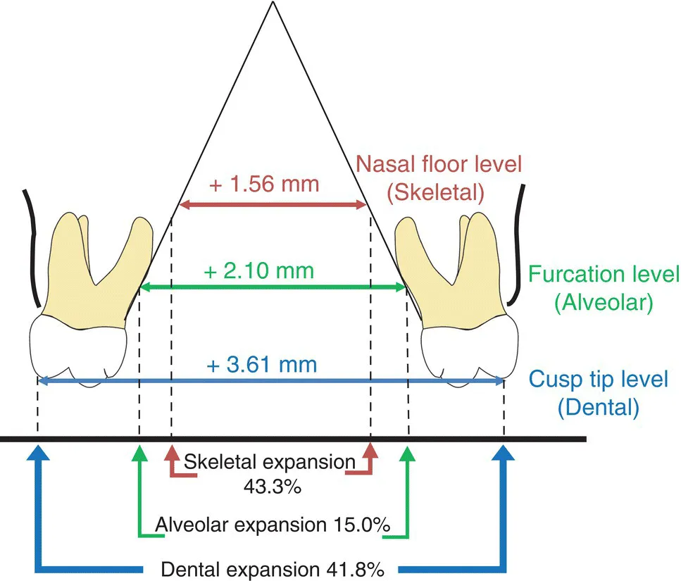
The result of maxillary expansion was stable approximately four years after the treatment [2]. Immediately after the maxillary expansion, the amount of total expansion was composed of 39.1% of skeletal expansion (at the nasal floor level), 7.1% of alveolar expansion (at the furcation level), and 53.8% of dental expansion (at the cusp tip level). As the expansion showed slight but clinically insignificant relapse during retention, the composition of total maxillary expansion was 43.3% of skeletal expansion, 15.0% of alveolar expansion, and 41.8% of dental expansion one year after the expansion (Figure 33.3) [3]. Serial cone‐beam computed tomography (CBCT) images exhibited 1.6 mm apical movement of the buccal alveolar crest of the maxillary first premolar one year after the expansion, while other periodontal measurements showed clinically insignificant changes during the same period. With regard to the nasal airway, the nasal cavity significantly increased in terms of cross‐sectional area and volume, while the nasopharynx did not show clinically significant changes [5].
Previous studies [15] have found that non‐surgical MARPE can be effectively used to correct maxillomandibular transverse discrepancy. The maxillary expansion by MARPE remained stable during the long‐term retention and may be helpful for cases of nasal obstruction by increasing the size of the nasal cavity [6]. However, clinicians should monitor periodontal changes carefully because of consequent buccal tipping of the posterior teeth followed by apical movement of the buccal alveolar crest.
Image described by caption.
Figure 33.1 Axial and coronal sections of the maxilla before (top) and after (bottom) MARPE (CBCT images, voxel size 0.3 mm). Note the midpalatal suture is open after the expansion (arrow).
Image described by caption.
Figure 33.2 The midsagittal image before (gray) and immediately after (blue). MARPE superimposed at the anterior cranial base (CBCT images, voxel size 0.3 mm). Note the anterior and inferior movement of the maxilla at the midsagittal section.
Image described by caption and surrounding text.
Figure 33.3 Schematic diagram of the maxillary expansion by MARPE. Eve...

Inhaltsverzeichnis