
eBook - ePub
Electromagnetic Analysis and Design in Magnetic Resonance Imaging
Jianming Jin
This is a test
Buch teilen
- 282 Seiten
- English
- ePUB (handyfreundlich)
- Über iOS und Android verfügbar
eBook - ePub
Electromagnetic Analysis and Design in Magnetic Resonance Imaging
Jianming Jin
Angaben zum Buch
Buchvorschau
Inhaltsverzeichnis
Quellenangaben
Über dieses Buch
This book presents a comprehensive treatment of electromagnetic analysis and design of three critical devices for an MRI system - the magnet, gradient coils, and radiofrequency (RF) coils. Electromagnetic Analysis and Design in Magnetic Resonance Imaging is unique in its detailed examination of the analysis and design of the hardware for an MRI system. It takes an engineering perspective to serve the many scientists and engineers in this rapidly expanding field.
Chapters present:
- an introduction to MRI
- basic concepts of electromagnetics, including Helmholtz and Maxwell coils, inductance calculation, and magnetic fields produced by special cylindrical and spherical surface currents
- principles for the analysis and design of gradient coils, including discrete wires and the target field method
- analysis of RF coils based on the equivalent lumped-circuit model as well as an analysis based on the integral equation formulation
- survey of special purpose RF coils
- analytical and numerical methods for the analysis of electromagnetic fields in biological objects
With the continued, active development of MRI instrumentation, Electromagnetic Analysis and Design in Magnetic Resonance Imaging presents an excellent, logically organized text - an indispensable resource for engineers, physicists, and graduate students working in the field of MRI.
Häufig gestellte Fragen
Wie kann ich mein Abo kündigen?
Gehe einfach zum Kontobereich in den Einstellungen und klicke auf „Abo kündigen“ – ganz einfach. Nachdem du gekündigt hast, bleibt deine Mitgliedschaft für den verbleibenden Abozeitraum, den du bereits bezahlt hast, aktiv. Mehr Informationen hier.
(Wie) Kann ich Bücher herunterladen?
Derzeit stehen all unsere auf Mobilgeräte reagierenden ePub-Bücher zum Download über die App zur Verfügung. Die meisten unserer PDFs stehen ebenfalls zum Download bereit; wir arbeiten daran, auch die übrigen PDFs zum Download anzubieten, bei denen dies aktuell noch nicht möglich ist. Weitere Informationen hier.
Welcher Unterschied besteht bei den Preisen zwischen den Aboplänen?
Mit beiden Aboplänen erhältst du vollen Zugang zur Bibliothek und allen Funktionen von Perlego. Die einzigen Unterschiede bestehen im Preis und dem Abozeitraum: Mit dem Jahresabo sparst du auf 12 Monate gerechnet im Vergleich zum Monatsabo rund 30 %.
Was ist Perlego?
Wir sind ein Online-Abodienst für Lehrbücher, bei dem du für weniger als den Preis eines einzelnen Buches pro Monat Zugang zu einer ganzen Online-Bibliothek erhältst. Mit über 1 Million Büchern zu über 1.000 verschiedenen Themen haben wir bestimmt alles, was du brauchst! Weitere Informationen hier.
Unterstützt Perlego Text-zu-Sprache?
Achte auf das Symbol zum Vorlesen in deinem nächsten Buch, um zu sehen, ob du es dir auch anhören kannst. Bei diesem Tool wird dir Text laut vorgelesen, wobei der Text beim Vorlesen auch grafisch hervorgehoben wird. Du kannst das Vorlesen jederzeit anhalten, beschleunigen und verlangsamen. Weitere Informationen hier.
Ist Electromagnetic Analysis and Design in Magnetic Resonance Imaging als Online-PDF/ePub verfügbar?
Ja, du hast Zugang zu Electromagnetic Analysis and Design in Magnetic Resonance Imaging von Jianming Jin im PDF- und/oder ePub-Format sowie zu anderen beliebten Büchern aus Medicine & Biotechnology in Medicine. Aus unserem Katalog stehen dir über 1 Million Bücher zur Verfügung.
Information
Chapter 1
Introduction to Magnetic Resonance Imaging
1.1 Introduction
Magnetic resonance imaging, commonly known as MRI, is a powerful non-invasive imaging technique that has played and will continue to play an important role in the medical community. In clinical practice, it can assist physicians in both diagnosis and presurgical planning with limited risk to the patient. In laboratory research, it can help neurologists and other biological scientists to discover novel basic anatomical structures and physiological principles. Unlike some other imaging techniques like x-ray computed tomography (CT), MRI does not require exposure of the subject to ionizing radiation and hence is considered safe. It also provides more information than other imaging modalities since MR signals are sensitive to several tissue parameters.
MRI belongs to a larger group of techniques which are based on the phenomenon of nuclear magnetic resonance (NMR). This phenomenon was discovered in bulk materials by Bloch and Purcell in 1946. As we will see later, certain atomic nuclei, when placed in a static magnetic field, will assume one of the two states: one has a higher energy level and the other has a lower energy level (Fig. 1.1). The energy difference between the two states is linearly proportional to the strength of the applied magnetic field. This is known as the Zeeman effect. In thermal equilibrium, the number of nuclei in the higher energy state is slightly less than the number of nuclei in the lower energy state. A nucleus in the higher energy state can fall to the lower energy state by emitting a photon with energy equal to the energy difference between the two states. A nucleus in the lower energy state can jump to the higher energy state by absorbing a photon with energy matching the energy difference between the two states. Therefore, when the nuclei in the applied magnetic field are irradiated by such photons, which are actually electromagnetic fields of certain frequency generated by a radiofrequency (RF) probe, some nuclei in the lower energy state will absorb the photons and jump to the higher energy state. This destroys the thermal equilibrium. Immediately after the irradiation of photons, the excess nuclei in the higher energy state will return to the lower energy state to recover the equilibrium, emitting photons or electromagnetic fields which can be detected by an RF probe. Since the frequency of the emitted electromagnetic signals is determined by the energy difference of the two states of the nuclei and the decay of the signals in time depends on the molecular environment of the nuclei, the NMR signals received by an RF probe can be analyzed to study the properties of the nuclei and their environment.
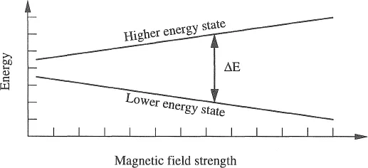
Figure 1.1. Splitting of energy level caused by the application of a static magnetic field.
For many years, NMR was primarily used for spectroscopic analysis before Lauterbur (1973) proposed using it for imaging purposes. The basic principle of using NMR for imaging is simple. Since the energy difference between the two states of certain nuclei in an external field depends on the strength of the external field, this energy difference at each point in the object to be imaged can be made different by varying the magnetic field from point to point. As a result, the energy of the photons and, consequently, the frequency of the electromagnetic fields absorbed or emitted by the nuclei are also different from point to point. After signals from all nuclei are received, their frequency may be used to determine spatial information about the nuclei. This simple phenomenon provides a basic foundation for all NMR imaging although the actual imaging methods are more complicated. The number of imaging techniques has blossomed since Lauterbur’s pioneering work. Technological developments have also increased the quality of NMR images. For example, the advent of superconducting magnets has made possible higher signal-to-noise ratio (SNR) and higher image resolution than were possible with resistive or permanent magnets. Modern computers have made possible whole volume imaging techniques which offer increased SNR and decreased image acquisition times. So-called “artifacts,” which are the distortions of the image due to unwanted effects such as motion, can be controlled using complex RF pulse sequences. Advances in coil technology have improved image quality and made techniques such as surface and microscopic imaging possible.
In this chapter, we first de...