
eBook - ePub
The Vulva
Physiology and Clinical Management, Second Edition
Miranda A. Farage, Howard I. Maibach, Miranda A. Farage, Howard I. Maibach
This is a test
Buch teilen
- 337 Seiten
- English
- ePUB (handyfreundlich)
- Über iOS und Android verfügbar
eBook - ePub
The Vulva
Physiology and Clinical Management, Second Edition
Miranda A. Farage, Howard I. Maibach, Miranda A. Farage, Howard I. Maibach
Angaben zum Buch
Buchvorschau
Inhaltsverzeichnis
Quellenangaben
Über dieses Buch
A unique compilation of expertise on anatomy, physiology, clinical issues, and current research, this textbook analyzes the range of presentation with age, ethnicity, symptoms, disorders, diagnoses, and toxicity. The second edition of this essential resource for anyone taking care of female patients has been doubled in scope to include additional chapters. All physicians, whether dermatologists or gynaecologists, as well as those researching the scientific evidence and symptoms, will benefit from the experience of the expert contributors and editors gathered here.
Häufig gestellte Fragen
Wie kann ich mein Abo kündigen?
Gehe einfach zum Kontobereich in den Einstellungen und klicke auf „Abo kündigen“ – ganz einfach. Nachdem du gekündigt hast, bleibt deine Mitgliedschaft für den verbleibenden Abozeitraum, den du bereits bezahlt hast, aktiv. Mehr Informationen hier.
(Wie) Kann ich Bücher herunterladen?
Derzeit stehen all unsere auf Mobilgeräte reagierenden ePub-Bücher zum Download über die App zur Verfügung. Die meisten unserer PDFs stehen ebenfalls zum Download bereit; wir arbeiten daran, auch die übrigen PDFs zum Download anzubieten, bei denen dies aktuell noch nicht möglich ist. Weitere Informationen hier.
Welcher Unterschied besteht bei den Preisen zwischen den Aboplänen?
Mit beiden Aboplänen erhältst du vollen Zugang zur Bibliothek und allen Funktionen von Perlego. Die einzigen Unterschiede bestehen im Preis und dem Abozeitraum: Mit dem Jahresabo sparst du auf 12 Monate gerechnet im Vergleich zum Monatsabo rund 30 %.
Was ist Perlego?
Wir sind ein Online-Abodienst für Lehrbücher, bei dem du für weniger als den Preis eines einzelnen Buches pro Monat Zugang zu einer ganzen Online-Bibliothek erhältst. Mit über 1 Million Büchern zu über 1.000 verschiedenen Themen haben wir bestimmt alles, was du brauchst! Weitere Informationen hier.
Unterstützt Perlego Text-zu-Sprache?
Achte auf das Symbol zum Vorlesen in deinem nächsten Buch, um zu sehen, ob du es dir auch anhören kannst. Bei diesem Tool wird dir Text laut vorgelesen, wobei der Text beim Vorlesen auch grafisch hervorgehoben wird. Du kannst das Vorlesen jederzeit anhalten, beschleunigen und verlangsamen. Weitere Informationen hier.
Ist The Vulva als Online-PDF/ePub verfügbar?
Ja, du hast Zugang zu The Vulva von Miranda A. Farage, Howard I. Maibach, Miranda A. Farage, Howard I. Maibach im PDF- und/oder ePub-Format sowie zu anderen beliebten Büchern aus Médecine & Dermatologie. Aus unserem Katalog stehen dir über 1 Million Bücher zur Verfügung.
Information
PART 1
Anatomy and Physiology
1
Anatomy of the vulva
INTRODUCTION
The vulva, or pudendum, is a collective term for the external female genital organs that are visible in the perineal area. Knowledge of the basic anatomy of the vulva is necessary in order to understand its physiology and appropriately recognize the wide spectrum of vulvar pathology. To achieve these goals, the vulvar embryology is first presented, before describing the anatomy of the vulva in women of reproductive age. Lifetime changes in the vulva from birth to adulthood are described in Chapter 3.
EMBRYOLOGY OF VULVA
Early in the fifth week of embryonic life, the cloaca is divided by the urorectal septum, which gives rise to the perineum. Folds of tissue form on either side of the cloaca: the anterior folds are urogenital and the posterior folds are anal. The anterior folds meet at the midline to form the genital tubercle. The genital tubercle enlarges. In the male embryo, under the influence of androgens, the genital tubercle becomes the penis; in the female embryo, growth slows and it becomes the clitoris. On either side of the tubercle, the urogenital folds form the labia minora. In the indifferent stage, the labioscrotal swellings develop on either side of the urogenital folds. In the male embryo, under the influence of androgens, they differentiate into the scrotum; in the female, lacking androgenic stimulation, they remain largely unfused to become the labia majora. The definitive urogenital sinus gives rise to the vaginal vestibule, into which the urethra, vagina, and greater vestibular glands open.
ANATOMY OF THE VULVA
The vulva consists of the mons pubis, the labia majora, the labia minora, the clitoris, the hymen, the vestibule of the vagina, the urethral orifice, Skene’s glands, Bartholin’s glands, and the vestibular bulbs (Figure 1.1).
Anatomy of the Vulva
The anterior and posterior boundaries of the vulva extend from the mons pubis to the anus, respectively; its lateral boundaries lie at the genitocrural folds. The vulvar epithelium exhibits regional differences in tissue structure based on embryonic derivation. The skin-bearing mons pubis, perineum, and labia are derived from the embryonic ectoderm. Vulvar skin, like skin at other sites, has a keratinized, stratified, squamous epithelial structure with hair follicles, sebaceous glands, and sweat glands. The thickness of the degree of keratinization of vulvar skin decreases progressively from the labia majora, over the clitoris, to the labia minora. The vulvar vestibule, derived from the embryonic endoderm, is nonkeratinized. Chapter 2 describes in detail the regional tissue structure of the vulva.
Mons Pubis
The mons pubis (mons Veneris) is the rounded eminence in front of the pubic symphysis, which is formed by a collection of adipose tissue beneath the integument. During puberty, it becomes covered with hair up to its junction with the abdominal wall. The hair pattern, or escutcheon, of most women is triangular. Genetic and racial differences produce a variety of normal hair patterns, with approximately one in four women having a modified escutcheon with a diamond pattern.
Labia Majora
The labia majora are a pair of prominent longitudinal, cutaneous folds of fibro-adipose tissue that are homologous to the scrotum in the male. The structures bear epidermal tissue resembling the dartos tunic of the scrotum, as well as adipose tissue, areolar tissue, blood vessels, nerves, and glands. The labia majora also include the terminal extension of the round ligament and, occasionally, a peritoneal diverticulum, the canal of Nuck.
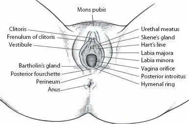
Figure 1.1 Anatomy of the adult vulva. (With kind permission from Libertas Academica Ltd, from Farage MA et al. Clin Med Womens Health 2010; 3: 1–13.)
The size of the labia majora is related to fat content. Each is approximately 7–8 cm in length and 2–3 cm in width. The labia majora extend downward and backward from the mons pubis, thus forming the lateral boundaries of a fissure or cleft (the pudendal cleft or rima) into which the vagina and urethra open.
Each labium majus has two surfaces: the outer surface is pigmented, rugose, and bears pubic hair, sebaceous glands, apocrine glands, and eccrine glands. The inner surface is smooth; it bears sebaceous, apocrine, and eccrine glands but no hair follicles. Vulvar apocrine glands are similar to those of the breast and axillary areas.
The labia majora are thicker in front. Anterior to the clitoris, they join to form the anterior boundary of the pudendal cleft, known as the anterior labial commissure. The labia majora do not surround the pudendal cleft fully; laterally, they remain approximately parallel to it and posteriorly, they gradually merge with the neighboring integument below the juncture of the labia minora (fourchette). The posterior ends of the labia majora and the connecting skin between them form the posterior boundary of the pudendum, known as the posterior labial commissure. The interval between the posterior commissure and the anus is 2.5–3 cm in length and constitutes the perineum.
Labia Minora
The labia minora (nymphae) are two small cutaneous folds that are situated between the labia majora and the vaginal orifice. The labia minora are homologous to the penile urethra and part of the skin of the penis in males. Laterally, they extend obliquely from the clitoris toward the rear for approximately 4 cm on either side of the vaginal orifice. They are shorter and thinner than the labia majora. At the clitoris, the anterior portion of each labium minus divides into two segments. Each upper segment passes anteriorly to the clitoris to meet its fellow of the opposite side, forming a fold, the preputium clitoridis, which overhangs the glans of the clitoris. Each lower segment passes beneath the clitoris, joining with its fellow to form the frenulum, which is attached to the inferior surface of the clitoris. The posterior portions of the labia minora surround the vestibule of the vagina. Their posterior juncture is the fourchette.
Histologically, the labia minora are composed of dense connective tissue, erectile tissue, and elastic fibers. Unlike the labia majora, they do not contain adipose tissue. The skin of the opposed surfaces of the labia minora has numerous sebaceous glands but no hair follicles or sweat glands. Among women of reproductive age, there is significant variation in the size of the labia minora. They are relatively more prominent in children and postmenopausal women.
Clitoris
The clitoris is a short, cylindrical, erectile structure that is 2–3 cm in length at the superior portion of the vestibule. It is the female homologue of the penis. It is situated beneath the anterior labial commissure, partially hidden between the anterior segments of the labia minora. The clitoris consists of a base of two crura that attach to the periosteum of the symphysis pubis. Like the penis, the clitoris has a suspensory ligament and two small muscles, the ischiocavernosi, which are inserted into the crura of the clitoris. The body of the clitoris consists of two cylindrical corpora cavernosa composed of thin-walled, vascular channels that function as erectile tissue. The distal third of the clitoris is a small rounded tubercle (glans clitoridis) that consists of spongy erectile tissue with many nerve endings. Usually, only the glans is visible, with the body of the clitoris positioned beneath the skin surface. The normal glans clitoridis in adult women has a width of less than 1 cm, with an average length of 1.5–2 cm. Age, weight, and oral contraceptive use do not change its anatomic dimensions. Childbearing may influence the size of the clitoris.
Hymen
The hymen is a thin fold of mucous membrane situated at the entrance to the vagina. Between the hymen and the frenulum of the labia minora is a shallow depression, the navicular fossa. The inner edges ...