Venous Ultrasound
eBook - ePub

Venous Ultrasound

Joseph A. Zygmunt Jr., Joseph A. Zygmunt Jr.

Buch teilen
  1. 229 Seiten
  2. English
  3. ePUB (handyfreundlich)
  4. Über iOS und Android verfügbar
eBook - ePub

Venous Ultrasound

Joseph A. Zygmunt Jr., Joseph A. Zygmunt Jr.

Angaben zum Buch
Buchvorschau
Inhaltsverzeichnis
Quellenangaben

Über dieses Buch

Venous Ultrasound 2e is the essential text for anyone involved in the treatment of chronic venous disease. It provides specific information on ultrasound as it is applied to chronic insufficiency, including history, general techniques, examples of anatomy, and protocols for performing ultrasound on patients, and discussions on key aspects of interpretation of sonographic findings.

Updated to include the outcome and impact of three recent studies, the ATTRACT trial, the EVRA study, and the VIDIO imaging trial. An entire chapter is dedicated to iliac venous and stent imaging for those interested in expanding practice based on the mentioned studies. Also included is specific protocol for imaging of the pelvic area with focus on the pelvic congestion and reflux affecting this anatomic area. This text demonstrates that as imaging techniques improve, so too will the understanding of venous pathologies increase and the burdens of their respective pathologies. Pelvic Congestion, iliofemoral and late stage disease can be interrogated with a non-invasive approach using the techniques included prior to interventional procedures.

This fully updated new edition includes coverage of new ablation techniques which include non- thermal and non- tumescent therapies for venous insufficiency – these have unique ultrasound properties on what to see, look for and observe in intra and post- operative situations.

Focusing on the fundamentals that every phlebologist needs to know, the color illustrations and numerous line drawings complement the text for a complete learning experience.

Key features:

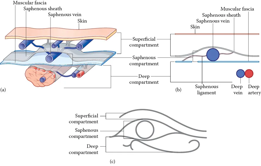
  • Covers anatomy related to venous insufficiency and obstruction
  • Protocols with step by step approaches for those new to certain exams
  • Includes useful diagrams and images to aid understanding
  • Thoroughly up to date, with all the latest information for those practicing venous therapies

Venous Ultrasound 2e is valuable for sonographers and physicians alike; including phlebologists, general and vascular surgeons, physicians, radiologists, angiologists, interventional cardiologist, mid-levels, and nurses who work in this area.

Häufig gestellte Fragen

Wie kann ich mein Abo kündigen?
Gehe einfach zum Kontobereich in den Einstellungen und klicke auf „Abo kündigen“ – ganz einfach. Nachdem du gekündigt hast, bleibt deine Mitgliedschaft für den verbleibenden Abozeitraum, den du bereits bezahlt hast, aktiv. Mehr Informationen hier.
(Wie) Kann ich Bücher herunterladen?
Derzeit stehen all unsere auf Mobilgeräte reagierenden ePub-Bücher zum Download über die App zur Verfügung. Die meisten unserer PDFs stehen ebenfalls zum Download bereit; wir arbeiten daran, auch die übrigen PDFs zum Download anzubieten, bei denen dies aktuell noch nicht möglich ist. Weitere Informationen hier.
Welcher Unterschied besteht bei den Preisen zwischen den Aboplänen?
Mit beiden Aboplänen erhältst du vollen Zugang zur Bibliothek und allen Funktionen von Perlego. Die einzigen Unterschiede bestehen im Preis und dem Abozeitraum: Mit dem Jahresabo sparst du auf 12 Monate gerechnet im Vergleich zum Monatsabo rund 30 %.
Was ist Perlego?
Wir sind ein Online-Abodienst für Lehrbücher, bei dem du für weniger als den Preis eines einzelnen Buches pro Monat Zugang zu einer ganzen Online-Bibliothek erhältst. Mit über 1 Million Büchern zu über 1.000 verschiedenen Themen haben wir bestimmt alles, was du brauchst! Weitere Informationen hier.
Unterstützt Perlego Text-zu-Sprache?
Achte auf das Symbol zum Vorlesen in deinem nächsten Buch, um zu sehen, ob du es dir auch anhören kannst. Bei diesem Tool wird dir Text laut vorgelesen, wobei der Text beim Vorlesen auch grafisch hervorgehoben wird. Du kannst das Vorlesen jederzeit anhalten, beschleunigen und verlangsamen. Weitere Informationen hier.
Ist Venous Ultrasound als Online-PDF/ePub verfügbar?
Ja, du hast Zugang zu Venous Ultrasound von Joseph A. Zygmunt Jr., Joseph A. Zygmunt Jr. im PDF- und/oder ePub-Format sowie zu anderen beliebten Büchern aus Médecine & Chirurgie et médecine chirurgicale. Aus unserem Katalog stehen dir über 1 Million Bücher zur Verfügung.

Information

Verlag
CRC Press
Jahr
2020
ISBN
9781000072266
1
Venous anatomy
Joseph A. Zygmunt Jr.
Introduction
Any discussion on venous anatomy has to begin with a history of man’s understanding of its form and function. In the second century, Galen related clear insights about venous disease and described interventions for varicose veins and venous ulcers. Although insights were noted, Galen’s physiology theory spoke of “natural spirits,” and that blood was created in the liver and somehow was attracted to the extremities [1].
Venous valves were first described in detail by Fabricius in Padua, Italy in 1579. However, it was not until the seventeenth century that a true understanding of venous flow was realized. Almost 400 years ago, in 1628, William Harvey, who was studying with Fabricius in Padua, first explained one-way circulation of blood in the body. Harvey, in his treatise du Mortu Cordis, described the contraction of the heart and the role of the veins in returning blood to the heart. Harvey also described the need for valves in the venous system to maintain the unidirectional aspects of blood flow. Given what Harvey taught us and the 400 years of science since, our understanding of the function and variability of the venous system continues to expand.
Our understanding of venous anatomy evolved significantly almost 30 years ago with the advent of duplex ultrasound. Steve Talbot introduced B-mode imaging in 1982, focused primarily on the deep system. This relatively simple to perform, noninvasive procedure allowed for detailed study. In the early 1990s, phlebologists began with earnest investigation of the superficial system. In 2004, renowned phlebologist Hugo Partsch said, “duplex ultrasound is the most significant contribution to the field of phlebology in the past ten years.” Current thinking of the key opinion leaders is that the venous system is actually more complex than the arterial system [2].
Consensus on nomenclature of anatomic terms
Duplex ultrasound has especially influenced our understanding of the anatomy of the superficial and deep venous systems of the lower extremities. In 2001, an International Interdisciplinary Committee met to update and refine the Terminologia Anatomica concerning the lower limb veins [3]. In 2005, a follow-up article with extensions, refinements, and clinical applications was published [4], setting the standards which are commonly employed in the international literature today. Some of the most notable revisions are encapsulated in Table 1.1.
Table 1.1 New nomenclature of key veins
New term
Old term
Femoral vein
Superficial femoral vein—eliminated
Great saphenous vein (GSV)
Long saphenous vein (LSV)—eliminated
Inguinal confluence
Saphenofemoral junction—still used
Posterior accessory of the GSV
Posterior arch vein-eliminated
Vein of Leonardo-eliminated
Small saphenous vein (SSV)
Lesser saphenous vein (LSV)—eliminated
Thigh extension of the SSV
Giacomini vein—still used
Posterior tibial perforating veins
Cockett vein—decreasing use
Most notable are the elimination of the term “superficial femoral vein,” which is actually part of the deep system, and elimination of the abbreviation of LSV and terms long or lesser saphenous vein as misleading and confusing, replaced by great saphenous vein (GSV) and small saphenous vein (SSV). Another significant delineation includes the concept of the muscular fascia as a boundary between the deep and superficial compartments, and the subdivision of the superficial compartment [58]. A basic depiction of the saphenous compartment is noted in Figure 1.1.
180829.webp
Figure 1.1 Depiction of the saphenous compartment (a,b) and Egyptian eye (c). (Reproduced with permission from Uhl JF and Gillot C. Phlebology 2007;22:194–206.)
The superficial compartment now includes the “saphenous subcompartment,” described as “duplication of the superficial fascia around the saphenous vein” and therefore always contains named saphenous vessels, and the “true superficial compartment,” which contains the epifascial tributaries. True duplication of the saphenous vein is now noted at only 2%. Further, the epifascial tributaries are typically responsible for the visible varicose veins noted on clinical examination. A few eponyms, such as “Giacomini vein,” “Cockett’s perforator,” and the term “posterior arch vein” are still used worldwide in the literature; however, most perforating veins are identified using anatomic location descriptors. Agreements on general terminology will be discussed, and specifics regarding the deep, superficial, and perforating vein systems will occur in their respective sections below.
General anatomic terms
The term peripheral is best used for the segment of the vein that is away from the heart; central, the segment of the vein toward the heart. With regard to a duplicated vein, this term is reserved for only when the two veins display the same path, topography, and relationships (like the tibial veins). If a vein is parallel but in a different compartment or plane, it cannot be considered doubl...

Inhaltsverzeichnis