
eBook - ePub
Medical Imaging Technology
Victor I. Mikla,Victor V. Mikla
This is a test
Buch teilen
- 154 Seiten
- English
- ePUB (handyfreundlich)
- Über iOS und Android verfügbar
eBook - ePub
Medical Imaging Technology
Victor I. Mikla,Victor V. Mikla
Angaben zum Buch
Buchvorschau
Inhaltsverzeichnis
Quellenangaben
Über dieses Buch
Medical Imaging Technology reveals the physical and materials principles of medical imaging and image processing, from how images are obtained to how they are used. It covers all aspects of image formation in modern imaging modalities and addresses the techniques, instrumentation, and advanced materials used in this rapidly changing field.
Covering conventional and modern medical imaging techniques, this book encompasses radiography, fluoroscopy, computed tomography, magnetic resonance imaging, ultrasound, and Raman spectroscopy in medicine. In addition to the physical principles of imaging techniques, the book also familiarizes you with the equipment and procedures used in diagnostic imaging.
- Addresses the techniques, instrumentation, and advanced materials used in medical imaging
- Provides practical insight into the skills, tools, and procedures used in diagnostic imaging
- Focuses on selenium imagers and chalcogenide glasses
Häufig gestellte Fragen
Wie kann ich mein Abo kündigen?
Gehe einfach zum Kontobereich in den Einstellungen und klicke auf „Abo kündigen“ – ganz einfach. Nachdem du gekündigt hast, bleibt deine Mitgliedschaft für den verbleibenden Abozeitraum, den du bereits bezahlt hast, aktiv. Mehr Informationen hier.
(Wie) Kann ich Bücher herunterladen?
Derzeit stehen all unsere auf Mobilgeräte reagierenden ePub-Bücher zum Download über die App zur Verfügung. Die meisten unserer PDFs stehen ebenfalls zum Download bereit; wir arbeiten daran, auch die übrigen PDFs zum Download anzubieten, bei denen dies aktuell noch nicht möglich ist. Weitere Informationen hier.
Welcher Unterschied besteht bei den Preisen zwischen den Aboplänen?
Mit beiden Aboplänen erhältst du vollen Zugang zur Bibliothek und allen Funktionen von Perlego. Die einzigen Unterschiede bestehen im Preis und dem Abozeitraum: Mit dem Jahresabo sparst du auf 12 Monate gerechnet im Vergleich zum Monatsabo rund 30 %.
Was ist Perlego?
Wir sind ein Online-Abodienst für Lehrbücher, bei dem du für weniger als den Preis eines einzelnen Buches pro Monat Zugang zu einer ganzen Online-Bibliothek erhältst. Mit über 1 Million Büchern zu über 1.000 verschiedenen Themen haben wir bestimmt alles, was du brauchst! Weitere Informationen hier.
Unterstützt Perlego Text-zu-Sprache?
Achte auf das Symbol zum Vorlesen in deinem nächsten Buch, um zu sehen, ob du es dir auch anhören kannst. Bei diesem Tool wird dir Text laut vorgelesen, wobei der Text beim Vorlesen auch grafisch hervorgehoben wird. Du kannst das Vorlesen jederzeit anhalten, beschleunigen und verlangsamen. Weitere Informationen hier.
Ist Medical Imaging Technology als Online-PDF/ePub verfügbar?
Ja, du hast Zugang zu Medical Imaging Technology von Victor I. Mikla,Victor V. Mikla im PDF- und/oder ePub-Format sowie zu anderen beliebten Büchern aus Technology & Engineering & Biomedical Science. Aus unserem Katalog stehen dir über 1 Million Bücher zur Verfügung.
Information
1
Advances in Imaging from the First X-Ray Images
X-rays as well as other forms of radiation continue to play a crucial role in medical and experimental science. X-rays are electromagnetic radiations having wavelengths roughly within the range from 0.05 to 100 Å. Their similarities to light led to the tests of established wave optics: polarization, diffraction, reflection, and refraction. Thanks the development of sophisticated computer technology, it is now possible to combine thousands of 2D X-ray slices into a 3D image for greater accuracy in pinpointing the location of tumors, bone breaks, and other internal ailments of the human body. X-rays are also widely used outside of the medical field. The wavelength of X-rays is comparable to the size of atoms. Therefore, they are ideally suited for probing the structural arrangement of atoms and molecules in a wide range of materials. The sections of this review are presented in a logical order beginning with the discovery of X-rays, followed by selected experimental details, with the final sections covering diagnostic radiology physics and other, nonmedical, applications. Roentgens story is widely known, but people who helped lay radiology’s foundations remain essentially anonymous, especially to younger physicists and physicians.
Keywords
X-ray; diffraction; radiology; chest radiography; fluoroscopy; mammography; exposure dosimetry
1.1 X-Rays—Early History
The true start of imaging, medical and nonmedical, as we consider it today was in 1895 when Roentgen discovered the X-ray in his Wurzburg laboratory on November 8. Almost immediately after Wilhelm Conrad Roentgen announced the discovery of the X-ray, imaging techniques based on the discovery were implemented all over the world. For example, chest radiography became one of those early applications, even though the equipment seems crude by comparison to that of today, and certainly there was no knowledge of the potential deleterious effects of the ionizing radiation of X-rays. Roentgen’s invention heralded the start of the modern era of medical imaging.
German physicist, a person who studies the relationship between matter and energy, Wilhelm Conrad Roentgen (1845–1923) discovered X-rays in 1895 while he was experimenting with electricity (Figure 1.1). Because he did not really understand what these rays were, he called them X-rays. By 1900, however, doctors were using X-rays to take pictures (called radiographs) of bones, which helped them treat injuries more effectively. In 1901, Roentgen was awarded the first Nobel Prize for physics for his discovery of this short-wave ray. He donated all the prize money to his university. Keeping his will the newly discovered rays were called just X-rays (“x” is usually used to denote the unknown entity). He refused to take out patent on this discovery because he wanted the entire humankind to receive the benefits of X-rays. He even forbade naming of these rays after him. The reader can find historical details that describe events leading up to and occurring around the time of Roentgen’s discovery in excellent articles by Linton [1], Assmus [2], and Mould [3], to say about few. Roentgen tested his new rays to define their properties and made numerous observations before writing a paper that was submitted for publication on December 28, 1895, to a local scientific publication [4]. Roentgens prestige in the community was such that his paper was accepted without review and published immediately [4].
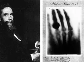
Figure 1.1 Wilhelm Conrad Roentgen (at the left) and Roentgen’s by-then-famous first X-ray image of his wife, Anna Bertha, hand (at the right). The X-rays penetrated through Anna’s skin, muscles, and other tissues but could not go through bones and wedding ring in her finger.
Scientific American published in its new section a short bulletin headlined “Professor Roentgen’s Wonderful Discovery” [5]. The first mention of the X-ray in Science magazine was a brief latter in the issue of January 31, 1896, from Hugo Munsterberg, a Harvard University physics professor [6]. Nature published a full translation of Roentgens paper [7]. Roentgen was dominated by a shyness that made him evade personal contacts wherever he could. In a brief period of time that follows, the X-ray went from a physics experiment to almost routine medical practice.
One can see what could be achieved in physics before the occurrence of the technological revolution involving not only computer applications but also the disappearance of the small independent X-ray companies into today’s multinational companies. Research and development is nowadays just too expensive for much independent practical high-technology contributions without financial backing.
1.2 The Use of X-Rays for Analysis
X-rays are a form of electromagnetic radiation, as is light (Figure 1.2). Their distinguishing feature is their extremely short wavelength—only about 1/10 000 that of light (or even less). This characteristic is responsible for the ability of X-rays to penetrate materials that usually absorb or reflect ordinary (visible) light.
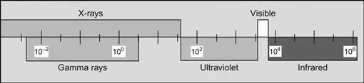
Figure 1.2 X-rays are part of the electromagnetic spectrum (wavelength is given in angstroms). X-rays have a wavelength in the range of 0.01–10 nm, corresponding to frequencies in the range of 3×10¹⁶–3×10¹⁹ Hz and energies in the range of 100 eV to 100 keV. They are shorter in wavelength than UV rays and longer than gamma rays.
X-rays exhibit all the properties inherent of light, but in such a different degree as to modify greatly their practical behavior. For example, light is refracted by glass and, consequently, is capable of being focused by a lens in such instruments as cameras, microscopes, telescopes, and spectacles. X-rays are also refracted, but to such a very slight degree that the most refined experiments are required to detect this phenomenon. Hence, it is impractical to focus X-rays. It would be possible to illustrate the other similarities between X-rays and light but, for the most part, the effects produced are so different—especially their penetration—that it seemed preferable to consider X-rays separately from other radiations. Figure 1.2 shows their location in the electromagnetic spectrum.
Their similarities to light led to the tests of established wave optics: polarization, diffraction, reflection, and refraction. Despite limited experimental facilities Roentgen could find no evidence of these. May be, it was additional reason he called them “x” (unknown) rays.
For diffraction applications, only short wavelength X-rays (hard X-rays) in the range of a few Angstroms to 0.1 Å (1−120 keV) are used. Because the wavelength of X-rays is comparable to the size of atoms, they are ideally suited for probing the structural arrangement of atoms and molecules in a wide range of materials. The energetic X-rays can penetrate deep into the materials and provide information about the bulk structure.
The year 2012 marks the 100th anniversary of the discovery of X-ray diffraction (XRD) and its use as a probe of the structure of matter [8–11]. Sixteen years after Rontgen announced in 1895 his discovery of “X” rays that can penetrate the body and photograph its bones, Max von Laue, a professor of physics at the University of Munich in Germany, worked on a theory of the interference of light in plane parallel plates. Laue was Plank’s favorite disciple. His interest covers the whole of physics. If, as some argued, X-rays were not made up of particles but were a form of electromagnetic radiation similar to ordinary (visible) light, and then it should be possible to repeat well-known optical experiments using X-rays instead of beams of ordinary light.
In 1911, Laue suggested to one of his research assistants, Walter Friedrich, and a doctoral student, Paul Knipping, that they try out X-rays on crystals. His reasoning was that X-rays have a wavelength close to the interatomic distances in crystals, and as a result, the crystal should act as a diffraction grating (Figure 1.3). Laue, always the theoretician, did not actually make the necessary experiments. In a single elegant experiment performed by Friedrich and Knipping, Laue had proven the wave-like nature of X-rays and the space–lattice structure of crystals at the same time [8,9]. When he received the Nobel Prize for what the Committee said was his “epoch-making discovery”, Laue gratefully acknowledged Friedrich and Knipping for their roles in the discovery; and for him it went without saying that he shared his prize money with them [10,11]. Einstein hailed Laue’s discovery as one of the most beautiful in the history of physics.
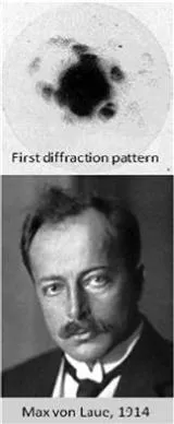
Figure 1.3 Max von Laue and the first diffraction pattern.
Before the Laue experiment, did anyone dream of tools allowing one to explore the structure of matter on the molecular scale and use this information for deriving structure–property relationships of materials—or for understanding the molecular basis of life? After X-rays had already been used to image the internal anatomy of the human body, with the Laue experiment the internal structure of crystals also became accessible on nanoscale [11].
Laue’s pioneering work in X-ray crystallography opened the way for two, quite different, developments in physics, both of them o...