
eBook - ePub
Fluorescent and Luminescent Probes for Biological Activity
A Practical Guide to Technology for Quantitative Real-Time Analysis
W. T. Mason, W. T. Mason
This is a test
Buch teilen
- 647 Seiten
- English
- ePUB (handyfreundlich)
- Über iOS und Android verfügbar
eBook - ePub
Fluorescent and Luminescent Probes for Biological Activity
A Practical Guide to Technology for Quantitative Real-Time Analysis
W. T. Mason, W. T. Mason
Angaben zum Buch
Buchvorschau
Inhaltsverzeichnis
Quellenangaben
Über dieses Buch
The use of fluorescent and luminescent probes to measure biological function has increased dramatically since publication of the First Edition due to their improved speed, safety, and power of analytical approach. This eagerly awaited Second Edition, also edited by Bill Mason, contains 19 new chapters and over two thirds new material, and is a must for all life scientists using optical probes.
The contents include discussion of new optical methodologies for detection of proteins, DNA and other molecules, as well as probes for ions, receptors, cellular components, and gene expression. Emerging and advanced technologies for probe detection such as confocal laser scanning microscopy are also covered. This book will be essential for those embarking on work in the field or using new methods to enhance their research. TOPICS COVERED:
* Single and multiphoton confocal microscopy
* Applications of green fluorescent protein and chemiluminescent reporters to gene expression studies
* Applications of new optical probes for imaging proteins in gels
* Probes and detection technologies for imaging membrane potential in live cells
* Use of optical probes to detect microorganisms
* Raman and confocal raman microspectroscopy
* Fluorescence lifetime imaging microscopy
* Digital CCD cameras and their application in biological microscopy
Häufig gestellte Fragen
Wie kann ich mein Abo kündigen?
Gehe einfach zum Kontobereich in den Einstellungen und klicke auf „Abo kündigen“ – ganz einfach. Nachdem du gekündigt hast, bleibt deine Mitgliedschaft für den verbleibenden Abozeitraum, den du bereits bezahlt hast, aktiv. Mehr Informationen hier.
(Wie) Kann ich Bücher herunterladen?
Derzeit stehen all unsere auf Mobilgeräte reagierenden ePub-Bücher zum Download über die App zur Verfügung. Die meisten unserer PDFs stehen ebenfalls zum Download bereit; wir arbeiten daran, auch die übrigen PDFs zum Download anzubieten, bei denen dies aktuell noch nicht möglich ist. Weitere Informationen hier.
Welcher Unterschied besteht bei den Preisen zwischen den Aboplänen?
Mit beiden Aboplänen erhältst du vollen Zugang zur Bibliothek und allen Funktionen von Perlego. Die einzigen Unterschiede bestehen im Preis und dem Abozeitraum: Mit dem Jahresabo sparst du auf 12 Monate gerechnet im Vergleich zum Monatsabo rund 30 %.
Was ist Perlego?
Wir sind ein Online-Abodienst für Lehrbücher, bei dem du für weniger als den Preis eines einzelnen Buches pro Monat Zugang zu einer ganzen Online-Bibliothek erhältst. Mit über 1 Million Büchern zu über 1.000 verschiedenen Themen haben wir bestimmt alles, was du brauchst! Weitere Informationen hier.
Unterstützt Perlego Text-zu-Sprache?
Achte auf das Symbol zum Vorlesen in deinem nächsten Buch, um zu sehen, ob du es dir auch anhören kannst. Bei diesem Tool wird dir Text laut vorgelesen, wobei der Text beim Vorlesen auch grafisch hervorgehoben wird. Du kannst das Vorlesen jederzeit anhalten, beschleunigen und verlangsamen. Weitere Informationen hier.
Ist Fluorescent and Luminescent Probes for Biological Activity als Online-PDF/ePub verfügbar?
Ja, du hast Zugang zu Fluorescent and Luminescent Probes for Biological Activity von W. T. Mason, W. T. Mason im PDF- und/oder ePub-Format sowie zu anderen beliebten Büchern aus Biowissenschaften & Biologie. Aus unserem Katalog stehen dir über 1 Million Bücher zur Verfügung.
Information
Part I
Introduction to Fluorescence Microscopy
CHAPTER ONE
Fluorescence Microscopy
JOHAN S. PLOEM, Medical Faculty, University of Leiden, The Netherlands
1.1 INTRODUCTION
1.1.1 Applications of fluorescence microscopy
As a tool in microscopy, fluorescence provides a number of possibilities in addition to absorption methods. Fluorescence probes can, for instance, be selectively excited and detected in a complex mixture of molecular species. It is also possible to observe a very small number of fluorescent molecules – approximately 50 molecules can be detected in 1 μm3 volume of a cell (Lansing Taylor et al., 1986). Furthermore, fluorescence microscopy offers excellent temporal resolution, since events that occur at a rate slower than about 10−8 s can be detected and measured with appropriate instrumentation. When confocal laser scanning is used in fluorescence microscopy, the theoretical limits of the spatial resolution (determined by the numerical aperture of the objective and the wavelength of the emitted fluorescence light) can be obtained in practice. In conventional microscopy, this is very difficult to obtain.
Immunofluorescence microscopy has been the most common application of fluorescence microscopy in cell biology (Coons et al., 1941). The possibility of detecting multiple regions, represented by specific antigens in the same cell, by selective binding of antibodies marked with fluorophores with different fluorescence colours is often used nowadays in in situ hybridization studies of, for example, DNA sequences in the interphase nucleus (Nederlof et al., 1990).
Fluorescence microscopy is also often used for the study of living cells (Kohen & Hirschberg, 1989). It is possible to measure, for example, the pH, free calcium and NAD(P)H concentration in the cytoplasm, as well as intercellular communications between cells. Flow cytometry as a specialized form of fluorescence microscopy (Melamed et al., 1990) permits the examination of biological surfaces when cells pass a beam of excitation light from a laser. A large number of cells can be analysed in a relatively short period of time by using several fluorescent probes in this technology.
1.1.2 The nature of fluorescence
Hot bodies that are self-luminous solely because of their high temperature are said to emit incandescence. All other forms of light emission are called luminescence. A system emitting luminescence is losing energy. Consequently, some form of energy must be applied from elsewhere and most kinds of luminescence are classified according to the source of this energy. One speaks, therefore, of electroluminescence, radioluminescence, chemiluminescence, bioluminescence and photoluminescence. In the latter form of luminescence the energy is provided by the absorption of ultraviolet, visible or infrared light. Fluorescence is a type of luminescence in which light is emitted from molecules for a very short period of time, following the absorption of light. The emitted light is termed fluorescence if the delay between absorption and emission of photons is of the order of 10−8 s or less. Delayed fluorescence is the term used if the delay is about 10−6 s, while a delay of greater than about 10−6 s results in phosphorescence. All these phenomena can be seen in microscopy.
1.1.3 Fluorescent stains
Compounds exhibiting fluorescence are called fluorophores or fluorochromes. When a fluorophore absorbs light, energy is taken up for the excitation of electrons to higher energy states. The process of absorption is rapid and is immediately followed by a return to lower energy states, which can be accompanied by emission of light. The spectral characteristics of a fluorochrome are related to the special electronic configurations of a molecule. Absorption and emission of light take place at different regions of the light spectrum (Fig. 1.1). According to Stokes’s law the wavelength of emission is almost always longer than the wavelength of excitation. It is this shift in wavelength that makes the observation of the emitted light in a fluorescence microscope possible. The excitation light of shorter wavelengths is prevented from entering the eyepieces by using the appropriate dichromatic (dichroic) dividing mirrors (Ploem, 1967). It should be noted that the intensity of the emitted light is weaker than that of the excitation light, as the emitted energy is much smaller than the energy needed for excitation. For different fluorochromes this may vary and is known as the quantum efficiency of the fluorophore used.
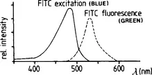
Figure 1.1 Excitation (absorption) and fluorescence (emission) spectra of fluorescein isothiocyanate (FITC).
Different fluorochromes are characterized by their absorption and emission spectra. The absorption or excitation spectrum is obtained by recording the relative fluorescence intensity at a certain wavelength when the specimen is excited with varying wavelengths. The most intense fluorescence occurs when the specimen is irradiated with wavelengths close to the peak of the excitation curve. An example of an absorption and an emission spectrum is given in Fig. 1.1. Most excitation and emission curves overlap to a certain extent.
Decrease in fluorescence during irradiation with light is called fading. The degree of fading depends on the intensity of the excitation light, the degree of absorption by the fluorophore of the exciting light and the exposure time (Patzelt, 1972). Reduction in fluorescence intensity can also be due to modification in the excited states of the fluorophore. These physicochemical changes may be caused by the presence of other fluorophores, oxidizing agents, or salts of heavy metals. This phenomenon is called quenching. Prior to microscopy a decrease in the potential to fluoresce can also occur. Preparations are therefore best stored in the dark at 4°C. To reduce fading during microscopy, agents such as DABCO (1,4-diazobicyclo-2,2,2-octane), N-propylgallate and p-phenylenediamine should be added to the mounting medium (Gilot & Sedat, 1982; Johnson & Nogueira Araujo, 1981).
1.1.4 Specialized literature on fluorescence microscopy
A number of books have been published recently on (quantitative) fluorescence microscopy and its applications. A few interesting examples are the books by Rost (1991), Kohen and Hirschberg (1989), and Lansing Taylor et al. (1986). Also, specialized techniques of fluorescence microscopy such as laser scanning fluorescence microscopy and confocal laser scanning microscopy have found wide applications, and consequently have been included in most recent books dealing with microscopy.
1.2 MICROSCOPE DESIGN
A fluorescence microscope is designed to provide an optimal collection of the fluorescence signal from the specimen, while minimizing the background illumination consisting of unwanted excitation light and autofluorescence. This requires rather sophisticated technology, since the specific fluorescence from the specimen can be several orders of magnitude weaker than the intensity of the exciting light. In the first place the fluorophore in the preparation must be excited with wavelengths as close as possible to the absorption peak of the fluorophore, assuming that the light source emits sufficiently in this wavelength region (Ploem, 1967). Secondly, the fluorescence emission collected by the optical system of the microscope must be maximized.
Strong excitation of the fluorophore with relatively efficient collection of the fluorescence is often not a good solution, since intense illumination may cause excessive fading of the fluorophore. Also, exciting light which does not correspond well with the excitation peak of the fluorochrome will often cause unnecessary autofluorescence of the tissue and optical parts, diminishing the image contrast. This contrast is determined by the ratio of the fluorescence emission of the specifically stained structures to the light observed in the background. For a good separation of exciting and fluorescence light the use of narrow-band excitation filters, which often have a relatively low transmission, is therefore necessary.
For easy visual observation, however, or photography with reasonably short exposure times, a sufficiently bright image is required. To that purpose a compromise between the intensity of the fluorescence and the level of background illumination must sometimes be accepted. If only a few fluorescent molecules are to be observed, not only the non-specific autofluorescence of tissue components, but also the level of autofluorescence of the glass components of the objective, immersion oil and the mounting medium can interfere with the observation of specific fluorescence. Laser scanning microscopy can provide a partial solution for these types of problems, as will be explained later in this chapter.
1.3 TYPESOFILLUMINATION
A fluorescence microscope is a conventional compound microscope. There are two basic types of illumination for fluorescence microscopy (FM): transmitted illumination (Young, 1961; Nairn, 1976) and incident illumination (Ploem, 1967; Kraft, 1973). The illumination pathway of transmitted light illumination is shown in Fig. 1.2. A condenser focuses the excitation light onto a microscope field. The emitted fluorescence is collected by the objective and observed through the eyepieces. In this configuration it is essential that two different lenses are used: a condenser to focus the excitation light on the specimen and an objective to collect the emitted fluorescence light. For optimal observation of fluorescing images these two lenses, which have independent optical axes, must be perfectly aligned. This i...