Principles of Nucleic Acid Structure
eBook - ePub

Principles of Nucleic Acid Structure

Stephen Neidle

Buch teilen
  1. 302 Seiten
  2. English
  3. ePUB (handyfreundlich)
  4. Über iOS und Android verfügbar
eBook - ePub

Principles of Nucleic Acid Structure

Stephen Neidle

Angaben zum Buch
Buchvorschau
Inhaltsverzeichnis
Quellenangaben

Über dieses Buch

This unique and practical resource provides the most complete and concise summary of underlying principles and approaches to studying nucleic acid structure, including discussion of x-ray crystallography, NMR, molecular modelling, and databases. Its focus is on a survey of structures especially important for biomedical research and pharmacological applications. To aid novices, Principles of Nucleic Acid Structure includes an introduction to technical lingo used to describe nucleic acid structure and conformations (roll, slide, twist, buckle, etc.). This completely updated edition features expanded coverage of the latest advances relevant to recognition of DNA and RNA by small molecules and proteins. In particular, the reader will find extensive new discussions on: RNA folding, ribosome structure and antibiotic interactions, DNA quadruplexes, DNA and RNA protein complexes, and short interfering RNA (siRNA). This handy guide ends with a complete list of resources, including relevant online databases and software.

  • Completely updated with expanded discussion of topics such as RNA folding, ribosome structure and antibiotic interactions, DNA quadruplexes, DNA and RNA protein complexes, and short interfering RNA (siRNA)
  • Includes a complete list of resources, including relevant online databases and software
  • Defines technical lingo for novices

Häufig gestellte Fragen

Wie kann ich mein Abo kündigen?
Gehe einfach zum Kontobereich in den Einstellungen und klicke auf „Abo kündigen“ – ganz einfach. Nachdem du gekündigt hast, bleibt deine Mitgliedschaft für den verbleibenden Abozeitraum, den du bereits bezahlt hast, aktiv. Mehr Informationen hier.
(Wie) Kann ich Bücher herunterladen?
Derzeit stehen all unsere auf Mobilgeräte reagierenden ePub-Bücher zum Download über die App zur Verfügung. Die meisten unserer PDFs stehen ebenfalls zum Download bereit; wir arbeiten daran, auch die übrigen PDFs zum Download anzubieten, bei denen dies aktuell noch nicht möglich ist. Weitere Informationen hier.
Welcher Unterschied besteht bei den Preisen zwischen den Aboplänen?
Mit beiden Aboplänen erhältst du vollen Zugang zur Bibliothek und allen Funktionen von Perlego. Die einzigen Unterschiede bestehen im Preis und dem Abozeitraum: Mit dem Jahresabo sparst du auf 12 Monate gerechnet im Vergleich zum Monatsabo rund 30 %.
Was ist Perlego?
Wir sind ein Online-Abodienst für Lehrbücher, bei dem du für weniger als den Preis eines einzelnen Buches pro Monat Zugang zu einer ganzen Online-Bibliothek erhältst. Mit über 1 Million Büchern zu über 1.000 verschiedenen Themen haben wir bestimmt alles, was du brauchst! Weitere Informationen hier.
Unterstützt Perlego Text-zu-Sprache?
Achte auf das Symbol zum Vorlesen in deinem nächsten Buch, um zu sehen, ob du es dir auch anhören kannst. Bei diesem Tool wird dir Text laut vorgelesen, wobei der Text beim Vorlesen auch grafisch hervorgehoben wird. Du kannst das Vorlesen jederzeit anhalten, beschleunigen und verlangsamen. Weitere Informationen hier.
Ist Principles of Nucleic Acid Structure als Online-PDF/ePub verfügbar?
Ja, du hast Zugang zu Principles of Nucleic Acid Structure von Stephen Neidle im PDF- und/oder ePub-Format sowie zu anderen beliebten Büchern aus Biowissenschaften & Molekularbiologie. Aus unserem Katalog stehen dir über 1 Million Bücher zur Verfügung.

Information

Jahr
2010
ISBN
9780080553528
1

Methods for Studying Nucleic Acid Structure

Publisher Summary

The advances in nucleic acid structural studies have been largely due to the increased power and sophistication of the experimental approach of X-ray crystallography, which have provided most of the highly detailed structural information to date. The dominance of the crystallographic approach still continues. Single-crystal X-ray crystallographic methods are able to determine the complete three-dimensional molecular structures of biological macromolecules without necessarily recourse to any preconceived model, provided the molecules are discrete and not the effectively infinite disordered polymers of nucleic acid fibers. Single crystals can be thus defined as ordered arrays of discrete and identical molecules in three dimensions. This approach is also useful for finding alternative crystal forms if initial trials produce crystals with poor diffraction or exceptionally large unit cells. The use of robotic crystallization methods is increasingly common. These enable rapid, large-scale screening of crystallization conditions to be undertaken, which is especially useful when dealing with difficult molecules such as large RNAs or protein-nucleic acid multisubunit complexes. Many nucleic acid crystallographers have developed specialized sets of conditions for their own speciality, notable examples being in the RNA and ribozyme field.

1.1 Introduction

Our knowledge of DNA and RNA three-dimensional structure has advanced immeasurably since the elucidation of the first such structure, that of the DNA double helix in 1953 by Watson and Crick in conjunction with the X-ray fiber diffraction data of Franklin and Wilkins. Fiber diffraction methods subsequently enabled the morphologies of a whole range of nucleic acid double helical types to become established. More recently, the relationships between DNA primary sequence and the fine details of its molecular structure have become increasingly understood, in large part from single-crystal and nuclear magnetic resonance (NMR) structural studies on defined-sequence oligonucleotides. DNA structure continues to surprise with its ability to exist in a wide variety of forms, such as left-handed and multiple-stranded helices. The study of RNA structure has a more recent history, which has revealed that RNA can fold in a wide variety of complex ways as well as occur in double-helical form. There is now a very large amount of experimental information on the structures of protein-DNA, protein-RNA, and drug-DNA complexes.
The discovery of the double helix, as Watson and Crick realized, immediately provided fundamental new insights into the nature of genetic events. We now have extensive knowledge of both the detail and the variety of DNA and RNA structures themselves, together with the manner in which they are recognized by regulatory, repair, and other proteins, as well as by small molecules. All this is giving us altogether more profound levels of understanding of the processes of gene regulation, transcription and translation, mutation/carcinogenesis, and drug action at the atomic and molecular levels. We are now beginning to piece together how all this works in the context of eukaryotic chromatin, so the challenges over the next few years will be to study the structural biology of large-scale DNA-protein structural assemblies just as has already been done for the ribosome.
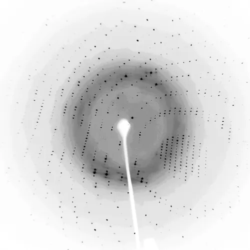
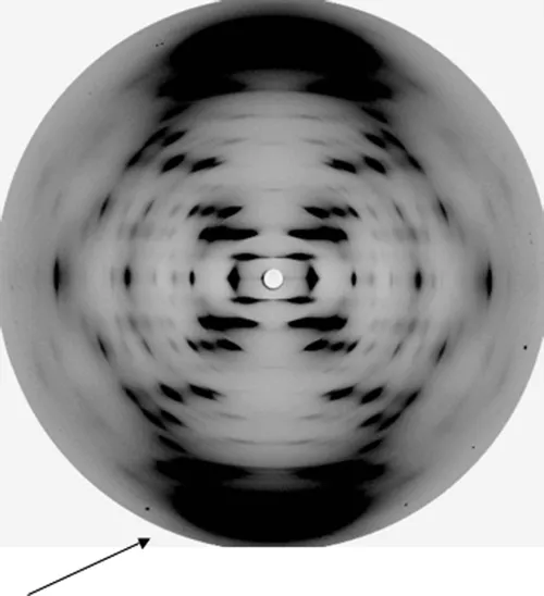
These advances in nucleic acid structural studies have been largely due to the increased power and sophistication of the experimental approach of X-ray crystallography, which have provided most of the highly detailed structural information to date. The dominance of the crystallographic approach still continues, and is reflected in the emphasis of this book. NMR spectroscopy, molecular modeling/simulation, and chemical/biochemical probe techniques also play important roles in providing information on structure, dynamics, and flexibility that can approach near-atomic resolution in at least some of its detail. Traditional spectroscopic-based biophysical methods can provide important complementary information, mostly at the macroscopic level. More recently developed techniques such as surface plasmon resonance spectroscopy and single-molecule methods, are extending their power so that the gap is now diminishing between macroscopic data on nucleic acids which the more traditional methods provide, and that at the atomic level. Underlying all of this progress have been the significant technical advances, notably in (i) the development of routine chemical methods for oligonucleotide synthesis and purification at the milligram level for both DNA and the more demanding RNA sequences, and (ii) the advent of efficient (and increasingly routine) cloning and expression systems for RNA and DNA-binding proteins, and for native RNA molecules.
This chapter provides a brief introduction to the two major structural methods, emphasizing their scope as well as their limitations for nucleic acid structural studies.

1.2 X-ray Diffraction Methods for Structural Analysis

1.2.1 Overview

X-rays typically have a wavelength of the same dimensions as interatomic bonds in molecules (about 1.5 Å). Scattering (or diffraction) of X-rays by molecules in ordered matter is the result of interactions between the radiation and the electron distribution of each component atom. Typical diffraction patterns from DNA, in the form of fibers or single crystals, are shown in Figs. 1.1, and 1.2. Reconstruction of the internal molecular arrangement by analysis of the scattered X-rays, analogous to a lens focusing scattered light from a microscope sample, thus provides a picture of the electron density distribution in the molecule. This reconstruction is not generally straightforward, due to the loss of phase information from the individual reflected X-rays during the diffraction process. The phase problem needs to be solved (see later for a brief description of various methods of doing this) in order for the electron density to be calculated in three dimensions (as a Fourier series), which is commonly termed a Fourier map. The approximate equivalence of the wavelength of X-rays and bond distance, of ~1–1.5 Å, means that in principle, the electron density of individual atoms in a molecule can be resolved, provided that the pattern of diffracted X-rays can be reconstituted into a real-space image.
image

Figure 1.1 X-ray diffraction pattern from a single crystal of a drug-oligonucleotide complex, taken with an image plate and a conventional laboratory X-ray source. The resolution limit for this pattern is 1.6 Å.
image

Figure 1.2 X-ray fiber-diffraction pattern from a sample of calf thymus DNA, showing a characteristic B-form pattern. (Provided by Professor Watson Fuller.) The arrow indicates the 3.4 Å layer line.
The degree of electron density detail that can actually be seen is dependent on the resolution of the recorded diffraction pattern. Resolution may be defined in terms of the shortest separation between objects (i.e. atoms or groups of atoms in a molecule) that can be observed in the electron density reconstituted from the diffraction pattern. The resolution limit (r) is governed by the maximum diffraction angle (θ) recorded for the diffraction data and the wavelength (λ) of the X-rays: r is defined as λ/2sinθ. At a resolution of 2.5 Å individual atoms in a structure cannot be resolved in an electron-density map although the shape and orientation of ring systems (e.g. base pairs) can be readily distinguished. These appear as elongated regions of electron density, with substituents being apparent as “outgrowths” from the main density. At 1.5 Å individual atoms are generally just about observable in a map, although only at about 1.0–1.2 Å are all atoms fully resolved and separated from each other (Fig. 1.3). There has been a marked increase in high-resolution studies (up to 0.8 Å) in recent years due to the increasing use and worldwide availability of high-flux synchrotron sources of X-rays for structural biology studies. There are currently about 20 synchrotrons with beam lines dedicated to crystallography, with several more scheduled over the next few years (see http://biosyn.rcsb.org for further details). In 2005, of the 4515 macromolecular crystal structures submitted to the Protein Data Bank (http://www.rcsb.org), data for 3398 (75.3%) were collected on synchrotron beam lines. The proportion in 2006 is even higher (78.1%). Synchrotron beam lines have intensities of X-ray beams greater by several orders of magnitude than conventional laboratory X-ray sources. Synchrotron facilities have also enabled much smaller crystals than previously to be successfully analyzed. Most importantly, the ability to tune X-rays to differing wavelengths has provided the means whereby powerful methods of structure analysis can be employed (see later). Although not comparable with synchrotron beam intensities, the development of highly effective mirror-optics focusing systems for laboratory X-ray sources in recent years can enable diffraction data to be collected in-house, especially when larger crystals can...

Inhaltsverzeichnis